Confocal Multi Photon Microscopy
Confocal microscopy scans the sample with a focussed light spot and allows for detection of fluorescence from thin sections also in thicker samples like eukaryotic cells. Two-photon excitation offers even further increased penetration depth compared to conventional confocal microscopy. Thereby bacterial behaviour in space and time can be followed directly in the host tissue.
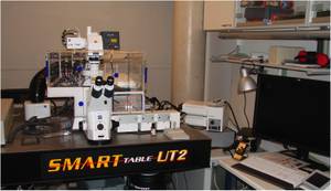
Zeiss LSM 710 NLO confocal microscope
Key features:
- multi photon: pulsed laser Mai:Tai BB (710 – 990 nm / <80 fs pulse width)
- confocal lasers: diode (405 nm) / argon (458/488/514 nm) / HeNe (561/633 nm)
- climate chamber
Research examples
Phenotypic heterogeneity and temporal expression of the capsular polysaccharide in Staphylococcus aureus
Shilpa George & Christiane Wolz
Department of Medical Microbiology
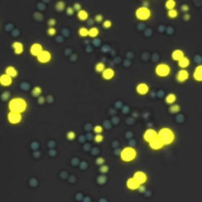
Bacteria respond to changing environments through adaptation. This leads to a high degree of phenotypic variability even in genetically homogeneous populations. In Staphylococcus aureus, the capsular polysaccharide protects against phagocytosis, but also impedes adherence to endothelial cells and matrix proteins. The regulation of the core biosynthesis genes (capA-P) required for capsular polysaccharide synthesis in S. aureus was studied at the single-cell level using promoter-fluorescent protein fusions. The regulatory molecule RNAIII required for capsule expression was analysed simultaneously. Pcap promoter activity appears yellow and PRNAIII activity blue. The quorum sensing effector molecule RNAIII is homogenously expressed in stationary cultures while the expression of the target gene capA is highly heterogeneous and expressed only in some cells.
George, S. E., Nguyen, T., Geiger, T., Weidenmaier, C., Lee, J. C., Liese, J. and Wolz, C. (2015), Phenotypic heterogeneity and temporal expression of the capsular polysaccharide in Staphylococcus aureus. Molecular Microbiology, 98: 1073–1088, doi: 10.1111/mmi.13174.
Staphylococcus aureus cells adhering to host cells
Christopher Weidenmaier Lab
Department of Medical Microbiology
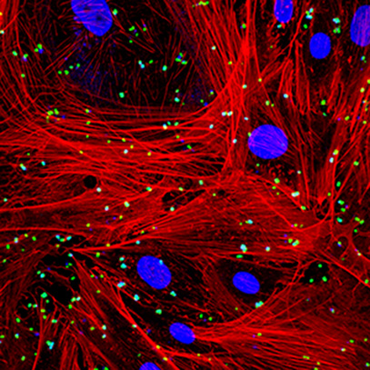
S. aureus cells labelled with a green fluorescent dye were brought into contact with host cells (nucleus stained blue and cytoskeleton red) under shear stress conditions in a flow chamber system. The flow chamber allows simulating conditions found e.g. in the blood stream or on epithelial surfaces in the nasal cavity. Bacteria adhering to the host cell surface and intracellular bacterial cells were monitored by confocal microscopy. Surface bound bacteria appear green while bacterial cells that have been taken up by the host cells lose their green fluorescence and appear blue or turquoise due to pH instability of the green dye. Using genetically engineered host and bacterial cells that exhibit altered receptors and surface structures the molecular mechanisms governing bacterial interaction with host cells are studied.