News
17.03.2023
Watching atherosclerosis as it develops
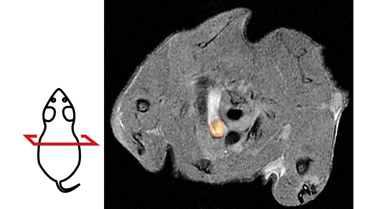
A team led by IFIB researcher Susanne Feil has developed a new method that can visualize diseased blood vessels. It may help to better understand heart attacks and strokes ‒ while reducing the use of laboratory animals.
The study was published in Circulation Research.
Publication:
Feil S, Stowbur D, Schörg B, Ehrlichmann W, Reischl G, Kneilling M, Pichler B, Feil R. 2023. Non-invasive detection of smooth muscle cell-derived hot spots to study atherosclerosis by PET/MRI in mice. Circ Res 132:747-750. DOI: 10.1161/CIRCRESAHA.122.322296
For further information, please see the press release.
Contact:
Dr. Susanne Feil
University of Tübingen
Interfaculty Institute of Biochemistry (IFIB)
Phone +49 7071 29-72458
susanne.feil@uni-tuebingen.de