Research

There is an ongoing need for high-quality binding reagents that provide insight into the structural conformation, binding partners, localization, and spatiotemporal dynamics of cellular components. Single-domain antibody fragments - referred to as nanobodies - derived from camelid heavy-chain antibodies have emerged as potent and versatile alternatives to conventional antibodies for numerous applications in biomedical research and therapy. Nanobodies are characterized by small size (2 -4 nm, ~ 14 kDa) and compact folding, resulting in high chemical stability, solubility and rapid tissue penetration. Due to their advantageous properties and accessibility to various cellular compartments, Nbs used as intrabodies have proven to be very suitable intracellular binding molecules.

Based on a long-standing expertise in recombinant antibody technologies, protein expression and nanobody generation, our research focuses on:

(a) Development, functionalization and application of nanobodies and functional derivatives thereof for crystallization studies, protein purification, analysis of protein-protein interactions, microscopy and flow cytometry as well as super-resolution microscopy and in vivo molecular imaging.

(b) generation of intracellular biosensors (chromobodies) to modulate, monitor and quantify components and processes in living cells using quantitative fluorescence microscopy.

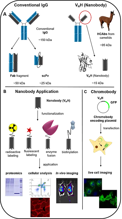
Fig.: Overview Nanobodies

A Schematic comparison between conventional IgG and heavy chain only antibodies (HCAbs) and derived recombinant antibody formats. While IgGs have two epitope binding domains (VH & VL) involved in antigen binding, HCAbs require only one variable domain (VHH). With a molecular weight of 15 kDa and a dimension of 2 x 4 nm, VHH is the smallest naturally occurring antigen binding moiety and thus also referred as “Nanobody” (Nbs).

B After selection of Nbs by in vitro display technologies, purified Nbs can be functionalized depending on the purpose, e.g. by radioactive or fluorescent labeling, coupling to an enzyme or biotinylation. This makes Nbs very versatile and reliable tools for a variety of applications in biomedical research.

C For visualization of endogenous antigens in live cells, Nbs can be genetically fused to fluorescent proteins and introduced as DNA-encoded expression constructs in living cells. Reflecting their chimeric structure these constructs are termed “chromobodies, CBs”. CBs are emerging tools to visualize and monitor spatiotemporal dynamics of endogenous target using quantitative live cell imaging.