Interfakultäres Institut für Mikrobiologie und Infektionsmedizin

Research - Team Oesterhelt

Influence of compatible solutes on the stability of membrane proteins

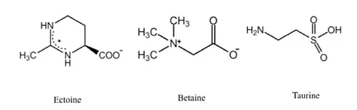
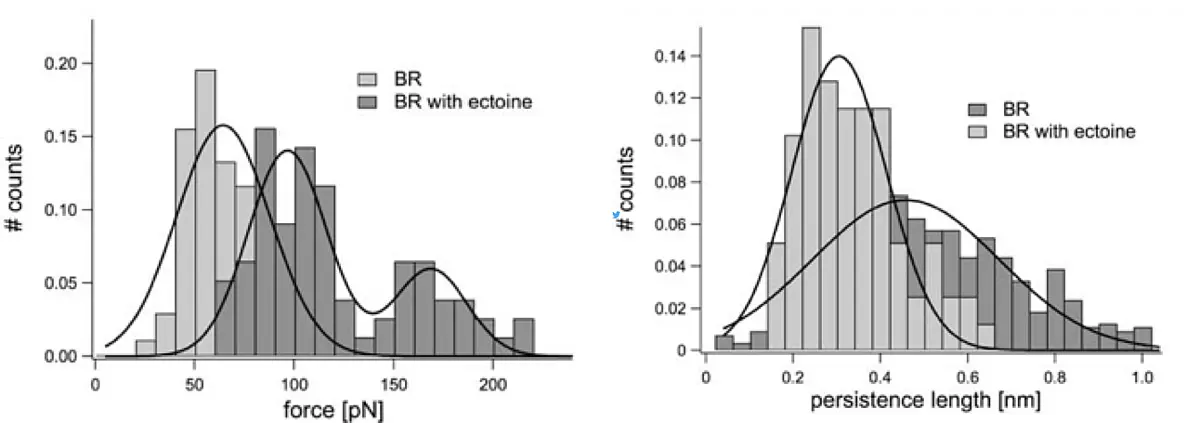
Compatible solutes are produced by microorganisms to protect cellular structures against environmental stresses like desiccation or high salt concentrations. They are small, zwitterionic, osmotically active substances, which can accumulate at high concentrations within bacterial cells, but even at molar concentrations they do not interfere with the cellular metabolism. Here, we studied the influence of different compatible solutes on the stability of the model membrane protein bacteriorhodopsin. Mechanical characteristics of bacteriorhodopsin were measured by single-molecule atomic force spectroscopy, applying different osmolytes at varying concentrations. In the presence of ectoin higher external forces were required to unfold bacteriorhodopsin, indicating overall protein stabilization. Also, the tendency of the unfolded amino acid strand to coil up was increased, demonstrating a higher tendency for protein refolding. In summary, the stabilizing influence of the compatible solutes on the structures of the membrane proteins was clearly observed, although the compatible solutes are known to establish no direct interactions with the proteins. The effect were observed for ectoin (...more) as well as for betain and taurin (...more).

Interactions of the transmembrane-helices in the light-harvesting complex II of Rhodospirillum photometricum

In the bacterial photosynthetic apparatus of R. photometricum the light-harvesting complex II (LH2) is located in the periphery and transduces the collected light energy to the central LH1 as well as the reaction centre. Although the crystal structure of LH2 had been solved, not much was known about the interactions occurring between the nine LH2 subunits within the LH2 ring. In collaboration with Simon Scheuring (INSERM/Univ. Aix-Marseilles) the combination of high-resolution force microscopy and force-spectroscopy was employed to study individual components of LH2 rings in native bacterial photosynthetic membranes. First, the LH2 rings were imaged within the membrane by force microscopy. Subsequently a single LH2 ring was manually unfolded and forces acting between individual subunits were measured by force spectroscopy. The occurrence of force peaks in periodic distances representing distinct unfolding events, indicated that unfolding occurred in distinct steps, some of which were coupled. About 150 pN were required to unfold an alpha-helix. Forces measured between the subunits of the LH2 ring indicated which subunits were in the process of interacting with each other. Interacting forces were rather weak, but sufficient to allow for self assembly of the subunits to the higher-order ring structure (...more).

Surface topology of Streptomyces spores

In collaboration with the group of Günther Muth (IMIT, Microbiology / Biotechnology) cover glasses with spore chains of Streptomyces sp. were imaged via an inverted optical microscope to select for hyphae at the specific stage of sporulation. The area of interest for AFM imaging was selected directly in an image of the spore chain taken by the CCD camera, using the "direct overlay" option provided in the AFM software.