Interfakultäres Institut für Mikrobiologie und Infektionsmedizin

Research - Team Brötz-Oesterhelt

Molecular Mode of Action of Antibiotic Acyldepsipeptides

Acyldepsipeptide antibiotics of the “ADEP” class, have potent antibiotic activity against a broad spectrum of multi-drug resistant Gram-positive bacteria in vitro including Methicillin-resistant Staphylococcus aureus (MRSA), Vancomycin-resistant Enterococcus sp. (VRE) and Penicillin-resistant Streptococcus pneumoniae (PRSP) (...more). ADEP treatment surpassed even the marketed antibiotic linezolid in lethal bacterial infections in rodents (...more). There is high medical need for treatment of bacteria persisting in the body in a state of dormancy, as they are a cause of recurrent infections and can hardly be cured by any antibiotic available. ADEP is exceptionally active against persister cells of S. aureus and E. faecium and outperformed all marketed antibiotics tested as comparators (work of Kim Lewis group, Northeastern University, Boston; ...more).

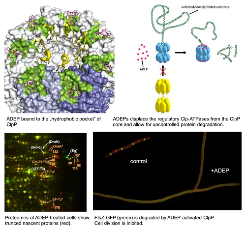
ADEP also stands apart from other antibiotics in terms of mode of action; it targets and dysregulates the proteolytic component of the bacterial Clp protease system, ClpP (...more).The bacterial Clp protease system plays an important role in maintaining protein homoeostasis within bacterial cells and in directing cellular differentiation and development programs. It is essential for bacterial virulence and serves as drug target of several recently discovered antibacterial natural products. ClpP is the conserved proteolytic core of the bacterial Clp protease complexes. For protein degradation, ClpP strictly depends on cognate Clp-ATPase partners. Without Clp-ATPases ClpP is only capable of digesting small peptides. Clp-ATPases act as a safety measure to ensure that ClpP degrades only such proteins that are destined for degradation (...more).

ADEP antibiotics unleash ClpP from these regulatory constrains. Upon binding of ADEP, ClpP sets out to degrade essential bacterial proteins, among those unstructured proteins and nascent polypeptide chains as they emerge from the ribosome. ADEP-treated cells die by self-digestion (...more). In firmicutes, such as staphylococci, streptococci, enterococci and bacilli the cell division regulator FtsZ proved to be especially sensitive to degradation by ADEP-activated ClpP and in these bacteria cell division inhibition is the primary cause of ADEP-mediated death (...more). FtsZ is also degraded in Wolbachia endobacteria, endosymbionts which pathogenic filarial worms require for reproduction (...more). In contrast, ADEP kills mycobacteria by inhibiting the indispensable natural functions of the Clp protease system (...more).

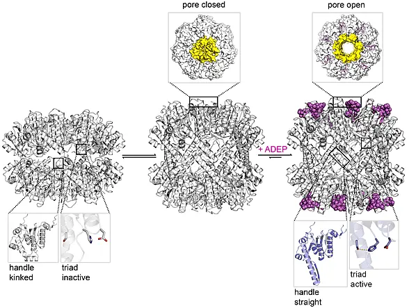
ADEP deregulates ClpP by extensive conformational control. ClpP is a 14-mer serine protease, with 14 catalytic centres shielded within its central proteolytic cavity. Small entrance pores prevent access of proteins to the proteolytic compartment. Upon binding of ADEP conformational rearrangements are set in motion. One consequence is that the gated pores widen, now allowing entry of proteins that the cell needs for survival. A second consequence is that ClpP is stabilised in the state where the catalytic triade adopts the conformation required for catalysis (...more). ADEP is the first antibiotic described to deregulate and over-activate its target; it turns a well-controlled bacterial protease into a deadly protein shredder.

Molecular basis of ADEP action. Binding of ADEP to the hydrophobic pocket triggers conformational changes in ClpP over a distance of 90 Angström. ClpP is stabilised in the extended conformation, the handle is stretched and forms contacts between the two heptameric rings. Only in this extended conformation the catalytic triad is in the correct distance to establish the hydrogen network required for catalysis. In addition, the entrance pores widen to allow for protein degradation (...more).

Molecular Mode of Action of Empedopeptin

Empedopeptin is an amphoteric, cyclic lipodepsipeptide antibiotic produced by the Gram-negative soil bacterium Empedobacter haloabium ATCC 31962 with antibacterial activity against a broad range of aerobic and anaerobic Gram-positive bacteria, including the important pathogens Staphylococcus aureus, Streptococcus pneumoniae and Clostridium difficile in vitro and animal models of bacterial infection. Despite these interesting features its specific mode of action had long remained elusive.

We found that empedopeptin interferes with the lipid cycle in peptidoglycan synthesis by sequestering lipid II as its primary target and additional lipid intermediates as secondary targets. Lipid II is a highly validated antibiotic target that is also addressed by the marketed antibiotic vancomycin. Enzymes processing these precursors are sterically inhibited and peptidoglycan synthesis comes to a halt. However, the binding site that empedopeptin recognizes at lipid II differs from that of vancomycin, with the advantage that empedopeptin remains active against vancomycin-resistant isolates. Empedopeptin binds lipid II in a region that involves at least the pyrophosphate group, the first sugar and the proximal parts of stem peptide and undecaprenyl chain. Calcium ions increase the antibacterial activity of empedopeptin by strengthening the interaction of the net-negative antibiotic with its target and with phospholipids in the cytoplasmic membrane (...more).

Mechanism of flavomannin B

The homodimeric flavomannin B and congeners were isolated from the endophytic fungus Talaromyces wortmannii. The compounds showed antibacterial activity against Staphylococcus aureus, including (multi)drug-resistant clinical isolates and relative selectivity for prokaryotes. Reporter gene analyses indicated induction of the SOS response, suggesting DNA strand breaks as a consequence of interference with DNA structure or metabolism. Accordingly, fluorescence microscopy demonstrated defective segregation of the bacterial chromosome and DNA degradation. The DNA gyrase inhibitor ciprofloxacin, which was tested for comparison, showed the same phenotype (...more).

Antibiotic Uptake across the Bacterial Cell Envelope

The medical need is particularly high for new agents against Gram-negative bacteria. The reason is limited compound penetration into Gram-negative cells, which are surrounded by two membranes with orthogonally different penetration requirements, the cytoplasmic membrane and the outer membrane. To better understand this phenomenon, we also study uptake routes of antibiotics across the bacterial cell envelope of Gram-negative bacteria. In order to determine to which extend the outer membrane forms a barrier for the entry of distinct antibacterial agents, Escherichia coli cells were grown with or without the addition of polymyxin B nonapeptide (PMBN). This compound, that represents the peptide portion of the antibiotic polymyxin B, impairs the integrity of the lipopolysaccharide layer composing the external leaflet of the outer membrane. The barrier function of the outer membrane is reduced. Our results show that PMBN affects the potency of the five selected model antibiotics to a different extend. While the minimal inhibitory concentrations (MICs) of tetracycline, doxycycline, and ampicillin dropped in the presence of PMBN, indicating increased potency and better uptake under these conditions, the MICs for cefoxitin and meropenem remained unchanged. In conclusion, uptake across the outer membrane is rate-limiting under these conditions for the first three but not for the latter two drugs.

Knockout studies using an isogenic set of porin mutants demonstrate, why the carbapenem meropenem can pass the outer membrane comparably easily: the compound can use multiple porins as entry routes. The porins OmpC and OmpF are particularly important and the porin LamB seems to form an additional entry route. Only if OmpC plus OmpF are deleted in parallel, significantly less meropenem reaches the periplasm, whereas single knock-outs of neither OmpC nor OmpF lead to a significant increase in the MIC. The single knock-out strains were obtained from the Keio collection, the multiple knockouts were prepared in our group using the same genetic background.