BiFC is a versatile technique for detection of protein-protein interactions in vivo (Hu et al., 2002). A fluorophore such as YFP is split into two non-fluorescing halves which are used to tag proteins of interest. Interaction of those proteins is monitored by assaying the fluorescence intensity. Absence of fluorescence is usually interpreted as a lack of interaction. BiFC however, has a couple of significant shortcomings that have not been addressed so far in a systematic way. Conventional BiFC lacks an internal reference marker; thus it is at best cumbersome to distinguish weak interactions from background fluorescence levels. Also, using two independent fusion proteins does not account for one being weaker expressed, resulting in misinterpretation of results, i.e. absence of or a seemingly weaker interaction does not necessarily reflect the true nature of that particular protein couple. Even Western data can not sufficiently exclude the possibility of differential expression in individual cells which are monitored for interaction (Xing et al., 2016).
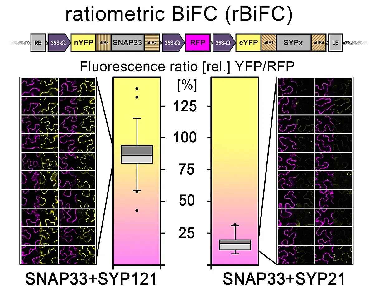
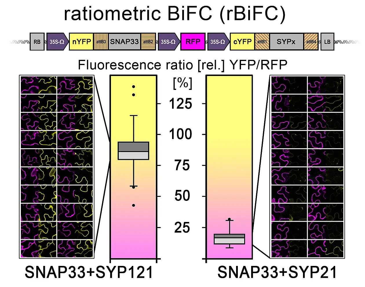
Through merging the BiFC with the novel “2in1” cloning system some previous issues of the technique will be avoided (Grefen and Blatt, 2012b). Being able to express both fusion proteins from the same T-DNA will allow expressing similar ratios (see figure 10, Hecker et al. 2015). In addition, a third expression cassette containing a fluorescent cytosolic marker (here: RFP) will act as an individual transformation control identifying cells that express the transgenes. This is particularly useful if a protein pair does not interact as each fusion construct alone is non-fluorescent. However, the major advantage of constitutive RFP expression is the possibility to quantify BiFC results. Through measuring mean fluorescent intensities of both fluorophores, a ratio can be calculated that will allow putting results from different protein pairs into context, grading different interaction strengths in a semi-quantitative approach. Additionally, myc and HA epitopes at the YFP-halves allow verification via immunoblot.