attempto online
08.02.2024
Core Facility for Electron Microscopy
Tübingen Structural Microscopy Core Facility (TSM) on the Morgenstelle Campus

Tübingen Structural Microscopy (TSM) is one of the University of Tübingen’s five Core Facilities funded by the German government’s Excellence Strategy. A total of eight electron microscopes are organizationally united under the umbrella of the Core Facility and are supervised by the participating laboratories of the Departments of Biology and Geosciences as well as the Center for Plant Molecular Biology (ZMBP).
"TSM's services include project consulting, support on the microscopes, and training on devices, techniques and methods. We assist research projects to varying degrees depending on the level of knowledge. This also includes methodological advice on submitting research proposals and writing publications," explains Dr. Stefan Fischer, Head of the Tübingen Structural Microscopy Core Facility (TSM). Fischer is a biologist and has been at the University of Tübingen since 2014. He was previously a postdoc in Halifax, Canada, where he specialized in 3D electron microscopy. TSM is involved in the training of young scientists in electron microscopy courses that are integrated into Bachelor's and Master's degree programs.
Along with Dr. Stefan Fischer, the TSM executive board consists of Professor Oliver Betz, group leader of Evolutionary Biology of Invertebrates at the Institute of Evolution and Ecology, Professor Andreas Kappler, Geomicrobiology group leader at the Department of Geosciences, and Dr. Mark Stahl, Head of Central Services at the Centre for Plant Molecular Biology (ZMBP). On the administrative side, Professor Peter Grathwohl, Vice-President of Research and Innovation, and Dr. Andrea Schaub, Head of the Research Division, are on the executive board.
Decentral but within walking distance
The TSM is organized decentrally. The member labs and electron microscopes are located on the Morgenstelle campus within a radius of less than 100 m between the Departments of Biology and Geosciences and the Center for Plant Molecular Biology (ZMBP). This means the available experience with sample preparation methods and the applications of electron microscopy is wide-ranging.
"What is particularly exciting is that the diversity of research topics and issues has increased greatly due to the departments involved. For new topics, we always have to specifically adapt and optimize processes, for example the fixation of samples. The quality of sample preparation is crucial for the quality of the subsequent analyses and their informative value. An important part of our work therefore lies in the consultation and design of electron microscopic investigations and the optimization of specific sample preparation", explains Stefan Fischer.
Coordination of new and existing electron microscopes
The Core Facility was founded in 2020 with the aim of pooling the electron microscopes already available on the Morgenstelle Campus which were not yet part of a Core Facility, and to be able to integrate new microscopes in the future.
"We operate two transmission electron microscopes (TEM), four scanning electron microscopes (SEM), a focused ion beam scanning electron microscope (FIB-SEM) and an electron probe microanalyzer (EPMA), which are located in our member labs in the Departments of Biology and Geosciences and in the Center for Plant Molecular Biology (ZMBP)." In addition to pure electron microscopy, the scientists also use a combination of fluorescence, confocal laser scanning and electron microscopy for their investigations.
"The Cryo-FIB-SEM installed at the Geo- and Environmental Center (GUZ) was added at the beginning of 2020 and is the first device to be centrally assigned to the TSM," says Stefan Fischer. "A special feature of this microscope is that, in addition to room-temperature samples, we can also examine frozen samples. This allows us to avoid chemical fixation processes and examine samples as close as possible to their native state." The device can not only image surfaces, but also ablate them with a gallium ion beam, which enables three-dimensional high-resolution examination methods.
Research projects in Geosciences and Biology
The TSM's equipment is currently being used primarily for research projects in the geosciences, biology and the Cluster of Excellence Controlling Microbes to Fight Infections (CMFI). As part of the CMFI, Prof. Dr. Lars Angenent and Dr. Bastian Molitor are currently performing electron microscopy investigations in the field of environmental biotechnology supported by the TSM. For example, a project on archaea, Methanothermobacter thermautotrophicus, was supported by electron microscopy. These play a major role in biotechnology as they produce methane, which can replace fossil natural gas. In addition, TSM is currently supporting a project on the formation and excretion of the iron-binding and antimicrobial organic compound pulcherrimin by Staphylococcus epidermis by Professor Andreas Kappler, Professor Andreas Peschel and Professor Heike Brötz-Oesterhelt.
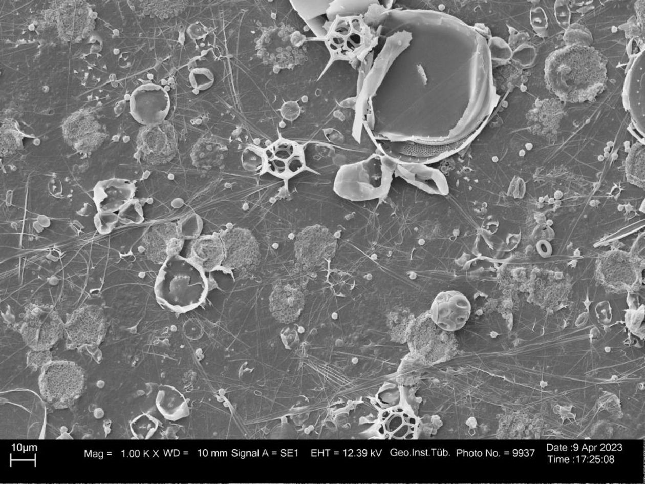
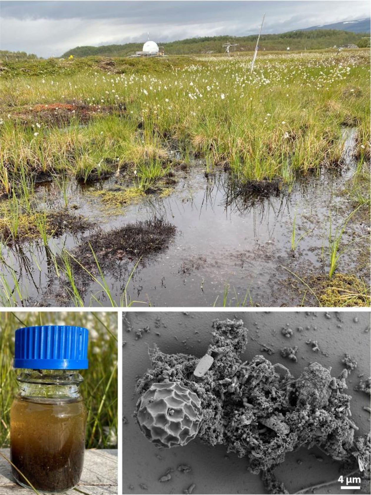
"In the geosciences, we have projects with environmental samples, for example, which aim to show the connection between the thawing of permafrost and the release of greenhouse gases (carbon dioxide and methane) by microorganisms," says Fischer. One project for which data was obtained at the TSM and which received greater media attention was the investigation of arsenic in soils and plants in rice fields in Asia by the Geomicrobiology working group led by Professor Andreas Kappler a few years ago.
Johannes Baral
