We have investigated metal-free organic magnets using a particular (and rarely accessible for small molecular compounds) microscopy.
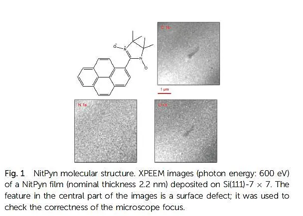
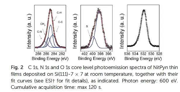
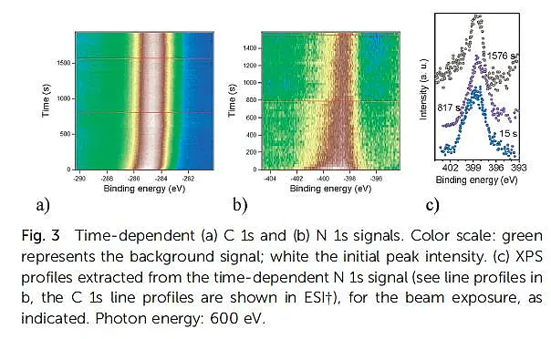
X-ray photoemission electron microscopy (XPEEM) with synchrotron radiation is a powerful microscopy technique that gives access to the lateral composition of interfaces. In combination with low energy electron microscopy, it offers the capability to correlate morphology, structure, and electronic properties. Our investigations are carried out with a spectroscopic photoemission and low-energy electron microscope (SPELEEM), installed at the Nanospectroscopy beamline, at Elettra Sincrotrone Trieste, which allows combining imaging, diffraction, and microprobe-spectroscopy. Microprobe-XPS is particularly useful due to the reduced acquisition times, which is vital in gaining information on the chemical bonding, without damaging the film. We presented a photoelectron emission microscopy investigation of the composition and stability of NitPyn, deposited on the Si(111)-7x7 surface. The strong interaction between the molecules and the surface influences not only the electronic structure of the molecule at the interface. Our results also suggest that the strong molecule-substrate interaction influences the growth mode of the thin films, favoring a layer-by-layer mode. We also investigated in real time the radiation sensitivity of NitPyn films. The observed degradation pattern, if successfully controlled and reproduced, can be exploited for its technological use in dosimeters and sensors.
The results are published in ChemComm in Open Access under the auspices of the University of Tübingen.
ChemComm 50 (2014) 13510
J Electron Spectrosc Relat Phenom (2015)