Interfaculty Institute of Microbiology and Infection Medicine

Research - Team Sass

Method platform to study bacterial cell division and its modulators

The divisome and elongasome are bacterial protein complexes responsible for peptidoglycan (PG) synthesis during cell division and elongation, respectively. The divisome is a highly dynamic macromolecular complex that is characterized by a time-dependent assembly of specific cell division proteins. Divisome formation is orchestrated by the tubulin homolog FtsZ. To initiate cell division, FtsZ assembles into protofilaments and forms a ring-like structure at the prospective division site. This “Z-ring” functions as a scaffold for the assembly of the bacterial cytokinetic machinery. Due to the increased emergence of bacterial resistance to established antibiotic classes, bacterial cell division has emerged as a promising new target pathway for antibiotic attack (...more). The group investigates the mechanism of cell division in bacteria and explores new approaches to target cell division with antimicrobial compounds.

With a focus on life-cell fluorescence microscopy and in vitro protein activity assays, we investigate the effect of antibiotics on different aspects of bacterial cell division. We either contribute to molecular mode of action elucidation of new antibiotics by following the consequences upon antibiotic treatment, or study fundamental principles of cell division using antimicrobial compounds as tools to selectively perturb the system. To test and characterize cell division inhibitors biochemically, we established in vitro assays to investigate the effect of such compounds on FtsZ from different species including Bacillus subtilis, Staphylococcus aureus, and Escherichia coli. For whole cell studies, we constructed a mutant library of fluorophore-tagged cell division proteins (single proteins/protein combinations) to investigate the consequences of antibiotics on cell division in whole cells using advanced microscopy techniques. For time-lapse and super-resolution fluorescence microscopy of living cells, we make use of the Nikon Eclipse Ti automated microscope equipped with a Perfect Focus system and the Zeiss Axio Observer Z1 automated microscope with LSM800 and AiryScan detector (see IMIT imaging platform).

ADEP inhibits cell division by activating bacterial ClpP to degrade FtsZ

The acyldepsipeptide antibiotic ADEP is a major compound under investigation. ADEP shows potent antibacterial activity against Gram-positive pathogens including streptococci, enterococci as well as multidrug-resistant Staphylococcus aureus (MRSA). ADEP acts by dysregulation of the caseinolytic protease Clp, a complex consisting of a proteolytic core, ClpP, that is flanked by corresponding Clp-ATPases. Protease activity of Clp is tightly controlled by Clp-ATPases that bind to distinct hydrophobic pockets of the barrel-shaped ClpP tetradecamer and thread the protein substrates into the degradation chamber. ADEP overcomes these strict control mechanisms by turning ClpP into an uncontrolled protease that now degrades flexible proteins like casein in the absence of Clp-ATPases, indeed leading to bacterial suicide. ADEP competes with and displaces the Clp-ATPases from ClpP, thereby preventing all its physiological functions. ADEP was the first antibiotic reported to kill bacteria by over-activating a non-essential target (...more). We showed recently that by binding to the hydrophobic pockets ADEP allosterically controls the conformation of ClpP locking its catalytic triads in an active conformation and opening the entrance pore to the degradation chamber (…more).

We identified the specific series of events from the physiological point of view that finally leads to death of ADEP-treated bacteria. Fluorescence microscopy and accompanying biochemical studies showed that ADEP prevents cell division in Gram-positive bacteria and induces filamentation of rod-shaped Bacillus subtilis and swelling of coccoid S. aureus and Streptococcus pneumoniae. ADEP treatment inhibits septum formation at the stage of Z-ring assembly, and central cell division proteins delocalize from mid-cell positions. This effect was due to the proteolysis of the essential cell division protein FtsZ which appeared to be particularly prone to degradation by ADEP-ClpP. Thus, ADEP inhibits a vital cellular process of bacteria that is not targeted by any therapeutically applied antibiotic so far. Therefore, their unique multi-faceted mechanism of action and antibacterial potency makes them promising lead structures for antibiotic drug discovery (...more).

Mycobacterial killing by ADEP is independent of FtsZ degradation

Many firmicutes like B. subtilis and S. aureus contain a single ClpP protein that is dispensable for normal growth. Here, the killing mechanism of ADEPs is based on activation of the ClpP protease to degrade essential proteins, i.e. FtsZ which is essential for bacterial cell division and thus survival of these species (see above). Recently, we found that the mechanism of growth inhibition can be different in species, where ClpP itself is essential for viability. Mycobacteria encode two ClpP homologs which are essential for growth, potentially due to the timely degradation of otherwise toxic proteins, making this organism an interesting candidate to further explore the effect of ADEP on actinobacteria. We observed that ADEP only weakly stimulated purified Mycobacterium tuberculosis ClpP1P2 to degrade larger peptides and unstructured proteins, and importantly, mtbFtsZ was not degraded during ADEP treatment and we did not detect significant inhibition of cell division during mycobacterial growth. In contrast to B. subtilis and S. aureus, mycobacteria are killed by a mechanism that does not dependent on the untimely degradation of FtsZ by ADEP-ClpP, but is caused by the inhibition of the natural functions of the essential mycobacterial Clp system (...more).

FtsZ is a phosphorylated by the Ser/Thr kinase PknB of S. aureus.

Synthesis of the bacterial cell envelope requires synchronization of a multitude of biosynthetic machineries and regulatory networks. The signal molecule sensed by the Ser/Thr kinase PknB of S. aureus, implicated in coordinating cross-wall formation, autolysis and cell division, remained elusive so far. Here, in collaboration with the labs of Fabian Grein and Tanja Schneider in Bonn, we could show that PknB interacts with lipid II, crosstalks with the two-component system WalKR, and phosphorylates the major cell division protein FtsZ. We observed PknB to preferentially localize to the septum triggered by its PASTA domains. The data provides a model for the role of PknB to control cell wall metabolism and cell division (...more).

Cell envelope stress sensor VraS is important for VISA-type resistance

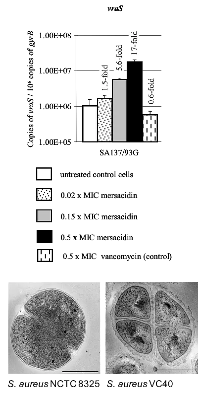
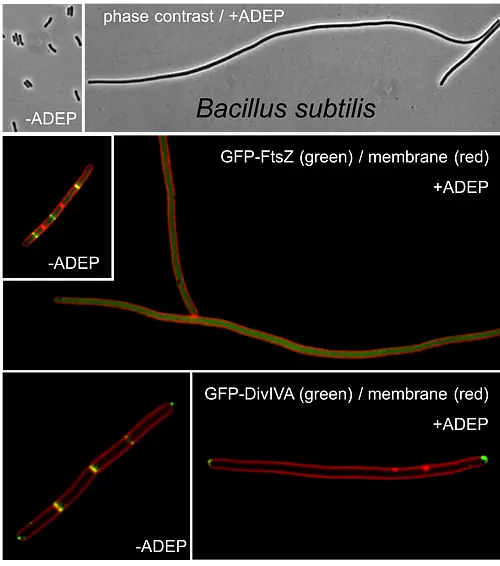
Bacterial cell wall stress response in S. aureus is mainly controlled by the two-component regulatory system VraSR that reacts to perturbation of cell wall synthesis by either cell wall-active substances, like vancomycin or daptomycin, or deregulated cell wall enzymes including MurF and Pbp2. The VraSR stimulon comprises the vraSR genes together with genes related to the cell wall metabolism of S. aureus like murZ, uppS, bacA, pbp2, sgtB and genes related to protein metabolism. We investigated the effect of the lantibiotic mersacidin, which acts by complexing the sugar phosphate head group of the peptidoglycan precursor lipid II thereby inhibiting the transglycosylation reaction of peptidoglycan biosynthesis, on inducing the cell wall stress response via VraSR. Our studies characterized the VraSR system as a stress sensing system (versus drug sensing system). We further found that mersacidin led to an extensive induction of the cell wall stress response, and in contrast to other cell wall-active antibiotics such as vancomycin, very low concentrations of mersacidin (0.15 x MIC) were sufficient for stimulon induction. The efficacy of mersacidin was not affected by an increased cell wall thickness, which is part of the VISA-type resistance mechanism and functions by trapping the vancomycin molecules in the cell wall before they reach lipid II. Therefore, the relatively higher concentration of mersacidin at the membrane might explain why mersacidin is such a strong inducer of VraSR compared to vancomycin (…more). In further studies, we characterized S. aureus VC40, a strain that shows full resistance to glycopeptides (vancomycin and teicoplanin MICs ≥32 mg/L) and daptomycin (MIC = 4 mg/L), and we could identify two amino acid exchanges in VraS amongst others by genome sequencing (…more). Transcriptomics indicated the increased expression of their respective regulons. Although not reaching the measured MIC for VC40, reconstitution of the L114S and D242G exchanges in VraS(VC40) into the susceptible parental background of S. aureus NCTC 8325 resulted in increased resistance to glycopeptides and daptomycin. The expression of VraS(VC40) led to increased transcription of the cell wall stress stimulon, a thickened cell wall, a decreased growth rate, reduced autolytic activity and increased resistance to lysostaphin-induced lysis in the generated mutant. Hence, a double mutation of a single gene locus, vraS, is sufficient to convert the vancomycin-susceptible strain S. aureus NCTC 8325 into a vancomycin-intermediate S. aureus (…more).

Phage endolysin to lyse bacterial cell wall and staphylococcal biofilms

Infections caused by Staphylococcus aureus and Staphylococcus epidermidis still play a major role in human and animal disease. In particular, staphylococcal biofilms on indwelling devices are difficult to treat due to their inherent antibiotic resistance. We investigated the possible use of phage endolysins to combat such a bacterial lifestyle. We cloned and heterologously overexpressed the lysis genes of the bacteriophages φ11 and φ12 of S. aureus NCTC8325 in Escherichia coli for subsequent analysis of the lytic activity of the full-length enzymes and their single subdomains on cell walls, whole cells, and staphylococcal biofilms. φ11 endolysin has been previously shown to possess a D-alanyl-glycyl endopeptidase and an N-acetylmuramyl-L-alanine amidase activity on crude cell walls of S. aureus OS2. While the φ12 endolysin was inactive and caused aggregation of the cells, we found that the φ11 endolysin hydrolyzed heat-killed staphylococci as well as staphylococcal biofilms. Cell wall targeting appeared to be a prerequisite for lysis of whole cells, and the combined action of the endopeptidase and amidase domains was necessary for maximum activity (…more).