Interfaculty Institute of Microbiology and Infection Medicine

Time-lapse Microscopy

Following the growth of individual bacterial cells over a prolonged period of time, from several minutes up to several hours or even days, provides unprecedented insight into their growth behaviour and morphological dynamics. By following the fate of fluorescently labelled cellular structures their arrangement in space and time can be studied. Such investigations require adjustable and stable conditions within the incubation chamber and high focus stability of the microscope.

Nikon Eclipse Ti-E inverted optical microscope

Key features:

Research examples

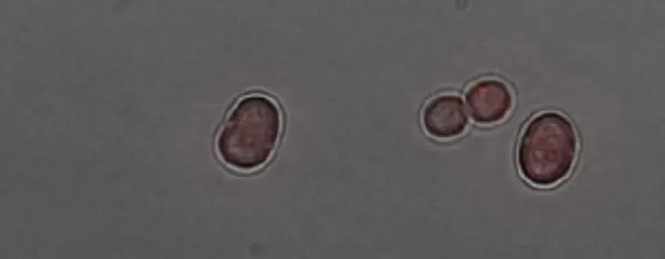
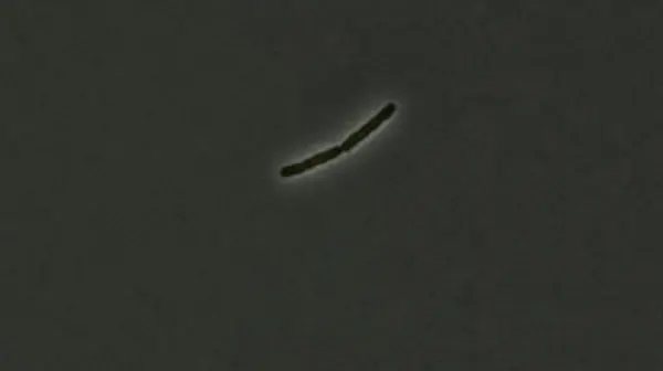
Germination of spore-like akinetes and heterocyst differentiation of filamentous cyanobacteria

Rebeca Perez1, Peter Sass2 & Iris Maldener1
Departments of Organismic Interactions1 and Microbial Bioactive Compounds2

Click here for the time-lapse video

Cyanobacteria are the bacterial ancestors of plant chloroplasts and perform oxygenic photosynthesis. Several strains grow in long filaments and differentiate into specialised cells, the heterocysts. These are the sites of nitrogen fixation and here nitrogen compounds are produced to supply the entire filament. Thereby, these bacteria are able to grow on any source of organic nutrition and to live on nothing but air and light. Cyanobacteria can be considered true multi-cellular organisms due to their division of labour between specialised cells with sophisticated cell-cell communication. Some strains form dormant spore-like akinetes to survive unfavourable conditions. Encountering beneficial conditions, they can germinate and differentiate into long filaments with heterocysts. Photosynthetically active cells are indicated by bright red autofluorescence.

Antibiotic-induced inhibition of cell division in Bacillus subtilis cells

Christian Mayer, Peter Sass & Heike Brötz-Oesterhelt
Department of Microbial Bioactive Compounds

Click here for time-lapse video

In most bacteria, including B. subtilis, bacterial cell division is coordinated by assembly of FtsZ as the pace maker protein at the division site. For initiating cell division, FtsZ localizes at mid-cell, the prospective site of septum placement, and forms a ring-like structure, the so-called FtsZ-ring (or Z-ring). The Z-ring functions as a scaffold for proteins that synthesize the division septum (“divisome”). A novel class of antibiotic acyldepsipeptides (ADEPs) prevents cell division in many Gram-positive bacteria and induces strong filamentation of rod-shaped B. subtilis cells. ADEP treatment inhibits septum formation at the stage of Z-ring assembly by activating a bacterial peptidase (ClpP) for the untimely degradation of FtsZ proteins. Here, time-lapse fluorescence microscopy allows following the delocalization of mCherry-tagged FtsZ upon ADEP treatment as well as the starting filamentous growth of the bacteria in real-time. In addition to mCherry-FtsZ (red), further fluorescently-labelled proteins like GFP-tagged Noc protein (green), which binds to specific noc sites in the oriC proximal two-thirds of the B. subtilis chromosome, can be simultaneously imaged to e.g. visualize unaffected, ongoing chromosome segregation in relation to FtsZ displacement.