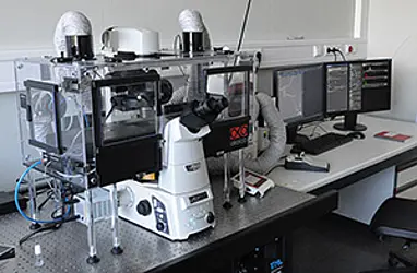
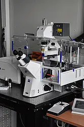
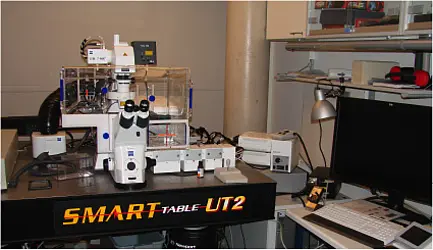
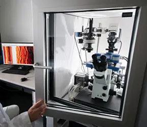
Three state-of-the art fluorescence microscopes tailored in their configurations to live-cell studies of bacteria are available in biosafety level II laboratories at the IMIT. These systems allow the IMIT research groups to study bacterial physiology, growth and development of pathogenic bacteria, bacteria-host cell interactions as well as the activity of antibacterial agents in real time and over prolonged periods.
The Nikon Eclipse Ti-E microscope is particularly suited for long-term fluorescence studies (“time-lapse studies”) due to its especially high focus stability. The Zeiss AiryScan microscope is based on a confocal set-up equipped with a newly developed light detection system and depicts growing cells with a resolution below the optical diffraction limit. The 2Photon microscope Zeiss LSM710 NLO resolves fluorescent bacteria embedded in host tissue and allows studies of the infection process.
In addition, an atomic force microscope (JPK NanoWizzard 3) is available, which scans surfaces with a sharp tip to reveal surface topographies with a resolution down to 10 nanometres.