Zentrum für Molekularbiologie der Pflanzen (ZMBP)

Research Group Jürgens

Membrane dynamics in cytokinesis


Plant cells divide by forming a cell plate which grows from the centre of the division plane to the periphery, eventually fusing with the plasma membrane. This mode of cell division is fundamentally different from the constriction of the plasma membrane and subsequent abscission of the midbody in non-plant cells. Cell-plate formation requires the orchestrated delivery of membrane vesicles along a dynamic cytoskeletal array called phragmoplast (Müller & Jürgens, 2016). These vesicles originate from the trans-Golgi network (TGN) and, after delivery to the plane of division, fuse with one another to initiate cell-plate formation and later with the margin of the centrifugally growing cell plate. 

            The fusion of membrane vesicles with one another or with a target membrane involves the formation of trans-SNARE complexes. Each SNARE complex comprises proteins with different SNARE domains (named Qa, Qb, Qc, R-SNARE); in some SNARE complexes a single Qbc-SNARE protein contributes two SNARE domains. The Qa-SNAREs (aka syntaxins) render SNARE complexes specific. In cytokinesis, the dominant Qa-SNARE is KNOLLE (KN, aka SYP111) whereas an evolutionarily ancient Qa-SNARE primarily involved in secretion (SYP132) also makes a contribution (Fig. 5; Park et al., 2018). The kn syp132 double mutant dies as a one-celled embryo with many nuclei. KNOLLE and SYP132 each interact with the same partners to form two types of SNARE complexes: (1) Qa KN (or Qa SYP132), Qb NPSN11, Qc SYP71, R VAMP721 (or VAMP722); (2) Qa KN (or Qa SYP132), Qbc SNAP33, R VAMP721 (or VAMP722) (El Kasmi and Krause et al., 2013; Park et al., 2018).

Figure 5. Evolution of cytokinetic SNARE complexes 

(A) Phylogenetic tree of SYP1 Qa-SNAREs in plant evolution

(B) With the advent of the angiosperms, cytokinesis-specific KNOLLE SNARE complexes were added to the ancient SYP132 SNARE complexes serving secretion and cytokinesis (Park et al., 2018).

           The KNOLLE-containing SNARE complexes already form at the ER membrane and are delivered as cis-SNARE complexes to the plane of cell division by membrane trafficking via Golgi stacks and TGN (Fig. 6; Karnahl & Park et al., 2017).

Figure 6Trafficking of cis-SNARE complexes during cytokinesis (model).

Two different types of cytokinetic cis-SNARE complexes are assembled on the ER, recruited into COPII vesicles and passed on to the Golgi stack/TGN. At the TGN, the cis-SNARE complexes are incorporated into AP1/CCV vesicles for delivery to the division plane. Following their disassembly by NSF ATPase, monomeric Qa-SNARE KNOLLE is assisted by SM protein KEULE in the formation of trans-SNARE complexes mediating fusion of adjacent vesicles during cell-plate formation and expansion (Karnahl & Park et al., 2017).

           Cell-plate formation involves newly-synthesised proteins including KNOLLE as well as proteins endocytosed from the plasma membrane which are recycled back to the plasma membrane in interphase. During cytokinesis, however, recycling of endocytosed PM proteins does not occur. These proteins are instead re-directed along a late-secretory pathway dependent on ARF-GEFs BIG1-4 to the plane of cell division (Richter et al., 2014).

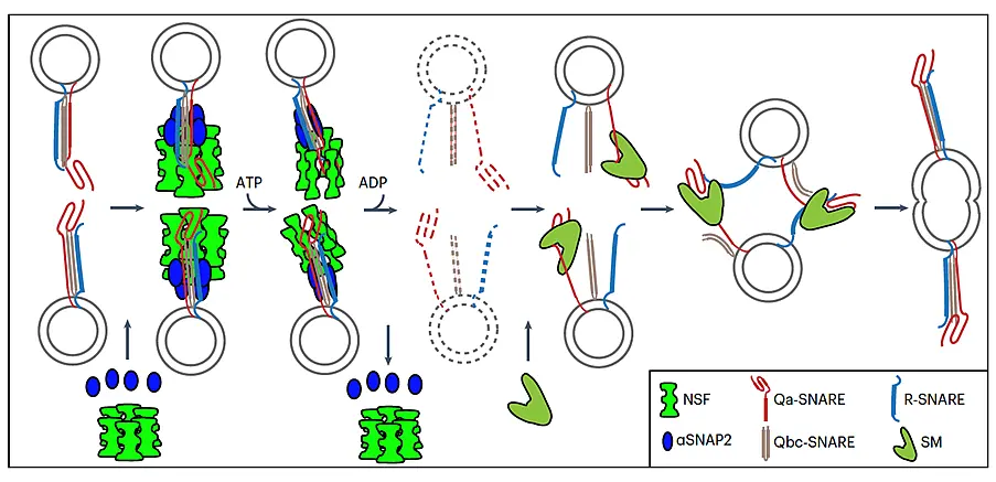
            Before vesicle fusion during cell-plate formation, the cis-SNARE complexes are disassembled by the action of AAA ATPase NSF and its co-factor aSNAP2 (Fig. 7; Park et al., 2023). Monomeric KNOLLE is then prevented from adopting an inactive "closed" state by interaction with the regulatory Sec1p/Munc18 (SM) protein KEULE (KEU), which promotes trans-SNARE complex formation between neighbouring membrane vesicles (Park et al., 2012).

Figure 7A mechanistic model of membrane fusion in Arabidopsis cell-plate formation involving disassembly of  cis-SNARE complexes by NSF ATPase and formation of trans-SNARE complexes promoted by SM protein KEULE

TGN-derived vesicles are delivered to the forming cell plate along the microtubules of the phragmoplast (not drawn). Hexameric NSF ATPase (bright green) and four copies of αSNAP2 adaptor (bright blue) form a 20S particle with the cis-SNARE complex (Qa-SNARE KNOLLE, red; Qbc-SNARE SNAP33, sand brown; R-SNARE VAMP721 or 722, dark blue) on each vesicle. ATP hydrolysis disassembles the cis-SNARE complex into monomeric SNAREs (broken lines, indicating a very transient state). The SM protein KEULE (pale green) interacts with monomeric fusion-competent KNOLLE. This interaction accelerates trans-SNARE complex formation between KNOLLE and its SNARE partners on adjacent vesicles, which mediates their fusion. (Park et al., 2023).

            Our current research addresses membrane dynamics in cytokinesis. To this end, we analyse how KN is targeted to its site of action and turned overhow KN interacts with other proteins, what cargo proteins are specifically delivered to the forming cell plate and how they act during cytokinesis.