Research projects
Biology of Streptomyces plasmids-Mechanism of conjugative DNA-transfer in antibiotic producing streptomycetes
Coworkers
Lina Thoma (PostDoc)
As producers of antibiotics, actinomycetes are regarded as the natural source and reservoir of antibiotic resistance genes which have been developed as part of the biosynthetic gene clusters to protect the producer from its own antibiotic. By horizontal gene transfer, the resistance determinants probably found their way into pathogenic bacteria causing major health problems.Streptomyces conjugation has been studied for 60 years but its underlying molecular mechanism is widely unknown. Conjugative DNA-transfer in mycelial actinomycetes is a unique process, differing considerably from conjugation via a type IV secretion system (T4SS), which has been well studied in many gram–negative and gram-positive bacteria (http://www.ncbi.nlm.nih.gov/pubmed/19946141).With few exceptions, Streptomyces plasmids do not encode any beneficial traits for the host cell. Even small Streptomyces plasmid, like pSVH1, (http://www.ncbi.nlm.nih.gov/pubmed/16439019) having a size of 8-15 kb and replicating via the rolling-circle mechanism are selftransmissible and encode all functions required for plasmid transfer and the mobilization of chromosomal markers.
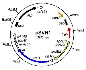
Fig.1 Map of the conjugative plasmid pSVH1 from the chloramphenicol producer Streptomyces venezuelae.
Functions involved in replication (black), regulation (red), transfer (blue) and intramycelial plasmid spreading (yellow) are indicated.
In contrast to conjugation via a T4SS, Streptomyces conjugation involves the transfer of a double-stranded DNA molecule (http://www.ncbi.nlm.nih.gov/pubmed/11679075). Under certain conditions, the Streptomyces DNA transfer process is associated with the formation of inhibition zones which have been named pock structures. Pock structures are formed, when spores of a plasmid carrying donor are plated with an excess of plasmid free recipient spores. Under these conditions pock structures of up to 3 mm develop in the growing mycelium.
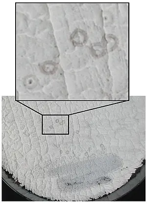
Fig. 2 Pock structures formed during conjugative transfer of plasmid pSVH1.
S. lividans TK23 carrying the pSVH1 derivative pEB211 was streaked on a lawn of plasmid free S. lividans TK64.
These “pocks” represent inhibition zones where growth and morphological differentiation is temporary retarded and indicate areas in the mycelial lawn where a recipient has obtained a plasmid by conjugation (https://www.ncbi.nlm.nih.gov/pubmed/27687731). Although the molecular mechanism underlying the formation of pock structures has not been elucidated, they have been interpreted as the consequence of intramycelial plasmid spreading within the recipient. This hypothesis has been recently confirmed by the fluorescence microscopic imaging of Streptomyces conjugation. Using a gfp-carrying derivative of the conjugative pIJ101 plasmid and a recipient strain encoding mCherry, donors, recipients and transconjugant mycelia could be differentiated. Observation of mating hyphae demonstrated that mating involves the lateral walls and not the primary tips, as it was previously suggested by the localization of a DNA-translocase TraB-GFP fusion protein to the hyphal tips (http://www.ncbi.nlm.nih.gov/pubmed/16776656). Since mCherry fluorescence was never detected in the donor compartments making contact to the recipient, it can be concluded that conjugative transfer does not involve intermixing of the cytoplasms, but a translocation pore specific for DNA-transfer (https://www.ncbi.nlm.nih.gov/pubmed/26286483). Following the initial transfer, the newly transferred plasmid spread from the contact sites of donor and recipient to the older parts of the recipient mycelium across septal cross walls.
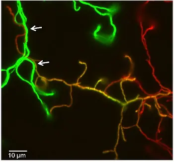
Fig. 3 Vizualization of conjugative transfer and plasmid spreading within recipient hyphae.
Spores of the donor S. lividans TK23 (pLT303) (green) and the recipient S. lividans T7–mCherry (red) were mixed and plated. After 20 h of growth at 29°C cells were imaged by fluorescence microscopy. Transconjugant hyphae appear yellow in the overlay. Scale bar, 5 µm.
A single plasmid encoded gene (traB) seems to be required for conjugative DNA-transfer (http://www.ncbi.nlm.nih.gov/pubmed/23082971). TraB proteins encoded by different Streptomyces plasmids are highly variable but resemble the septal chromosome translocator proteins FtsK/SpoIIIE in domain architecture, structure, and mode of DNA interaction. TraB of plasmid pSVH1 was shown to assemble to a hexameric ring-shaped structure that is able to form pores in planar lipid bilayers.
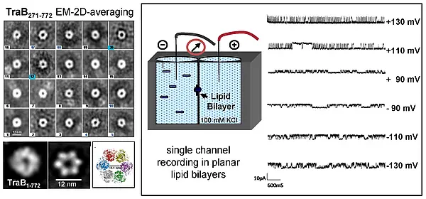
Fig. 4 Structure and pore forming ability of TraBpSVH1.
Electron microscopy of TraB shows a ring-shaped morphology. 2D averaging of electron microscopic images and homology modelling (template DNA-translocase domain of P. aeruginosa FtsK) of soluble TraB lacking the N-terminal membrane domain show a hexameric structure with a ~ 12 nm central channel. Single channel recordings in planar lipid bilayers revealed a pore forming ability of TraB.
TraB proteins bind to the plasmid localized cis acting locus of transfer (clt) by recognizing series of direct 8 bp repeats (TraB Recognition Sequence, TRS). Construction of various chimeric TraB proteins demonstrated that helix α3 of the c-terminal winged-helix-turn-helix (wHTH) fold determined sequence specific DNA recognition (http://www.ncbi.nlm.nih.gov/pubmed/21505418). clt-like sequences, which are also recognized by TraB are frequently detected in Streptomyces chromosomes (http://www.landesbioscience.com/journals/mge/11SepulvedaMGE1-3.pdf), but are absent or very rare in other actinomycetes (https://www.ncbi.nlm.nih.gov/pubmed/27687731). This suggests that mobilization of chromosomal markers is mediated by the interaction of TraB with the clt-like sequences and does not rely on the previous integration of the plasmid into the chromosome, as it is the case in the T4SS conjugation system (http://www.ncbi.nlm.nih.gov/pubmed/23082971).
The mechanism of intramycelial spreading via vegetative cross walls is still unclear. Depending on the conjugative plasmid, plasmid spreading requires 3 – 5 spd genes which do not possess any sequence similarity to functionally characterized proteins. Inactivation of a single spd gene abolishes plasmid spreading across the vegetative cross walls. The Spd proteins show multiple interactions with each other and with TraB, suggesting a multi-protein DNA-translocation apparatus involved in intramycelial plasmid spreading. From the eleven pSVH1 encoded proteins, six have a DNA-binding activity. The DNA-translocase TraB, the GntR-type transcriptional regulator TraR, and the replication initiator protein Rep recognize a specific target sequence (https://www.ncbi.nlm.nih.gov/pubmed/26015502). Also SpdA, involved in stable maintenance, binds to the spdA-associated palindromic DNA sequence (sps) highly conserved in many Streptomyces plasmids (http://www.ncbi.nlm.nih.gov/pubmed/25295034).
The characterization of the pSVH1 encoded Tra and Spd proteins suggested the following model of the Streptomyces conjugative DNA-translocation system (http://www.ncbi.nlm.nih.gov/pubmed/25592263). Streptomyces plasmids do not seem to encode a system for establishing mating pairs. Therefore, conjugative DNA transfer occurs only on solid agar during growth as substrate mycelium. Formation of multiple branching hyphae by apical tip extension provides good probability that mating partners will meet. Since Streptomyces hyphae grow by apical tip extension, the machinery for peptidoglycan remodeling is present at the hyphal tip and at new branching points. Presence of TraB probably directs fusion of the walls of the mating partners, maybe by interaction with a chromosomally encoded lytic transglycosylase.
After fusion of the PG-layer, TraB might also direct fusion of the membranes of donor and recipient as has been shown for SpoIIIE during sporulation of B. subtilis (http://www.ncbi.nlm.nih.gov/pubmed/16430687). Membrane insertion of TraB hexamers that assembled at clt sequences provides a pore structure for the DNA translocation.
TraB was shown to bind to the clt locus non-covalently without processing the plasmid (http://www.ncbi.nlm.nih.gov/pubmed/16776656). This raises the question, how TraB is able to translocate a circular DNA molecule. While during cell division the chromosome is already present at the closing septum, allowing FtsK to assemble at KOPS sequences at each chromosomal arm, TraB has to translocate the DNA across an intact membrane. Since it is highly unlikely that a circular molecule can be transported across the membrane, involvement of further DNA processing enzymes, e.g. a topoisomerase has to be postulated. Another possibility would be the fusion of the two TraB hexamers to a single channel structure releasing the circular plasmid end into the recipient.
In the recipient a vegetative cross wall traversing multi-protein complex consisting of Spd proteins is formed. By interaction with SpdB2 and other Spd proteins (http://www.ncbi.nlm.nih.gov/pubmed/17768240, www.ncbi.nlm.nih.gov/pubmed/27687731) the DNA translocase TraB is redirected to the septal crosswall and promotes plasmid transfer to the neighboring mycelial compartments.
Vector plasmids
Actinomycetes proteins are often difficult to express and purify in active form in E. coli. A series of stable replicating bifunctional expression vectors has been constructed to facilitate expression of proteins in Streptomyces strains.Most vectors are derived from the multi-copy, temperature-sensitive Streptomyces ghanaensis plasmid pSG5 replicating via a rolling-circle replication mechanism (RCR) and carry the pMB1 replicon for replication in E. coli. Resistance genes against thiostrepton (tsr) and kanamycin (aphII) or apramycin (aac(3)IV) allow selection in Streptomyces and E. coli.
Characteristics of some bifunctional Streptomyces (expression) vectors
vector | characteristics |
pGM190 | tsr, aphII, to terminator PtipA, RBS, fd terminator, sso, repts |
pGM191.1N | tsr, aphII, to terminator PtipA, RBS, N-terminal Strep-tagII-eGFP, fd terminator, sso, repts |
pGM202 | tsr, aphII, to terminator PtipA, RBS, C-terminal His6-tag, fd terminator, sso, repts |
pGM202-T7 | tsr, aphII, PT7, RBS,N- and C-terminal His-tags, fd terminator, sso, repts |
pGM202-T7-mcherry | tsr, aphII, PT7, RBS, mcherry, fd terminator, sso, repts |
pGM1190 | tsr, aac(3)IV, oriT, to terminator PtipA, RBS, fd terminator, sso, repts |
pGM1202 | tsr, aac(3)IV, oriT, to terminator PtipA, RBS, C-terminal His6-tag, fd terminator, sso, repts |
pGM-Gus | aac(3)IV, oriT, PermE_gusA, repts (knock out vector) |
pGus21 | aac(3)IV, oriT, PermE_gusA, oripMB1 (knock out vector, non-replicative in Streptomyces) |
Stable replication in Streptomyces was achieved by placing all genes in the same orientation as rep and by inserting the minus orgin sso (single stranded origin) for lagging strand replication. Inducible gene expression is mediated by either the PtipA promoter or the PT7 promoter (for use with S. lividans T7, constructed by J. Altenbuchner). Some, like pGM1202 carry the oriT fragment of plasmid RP4 and can be mobilized to streptomycetes by intergeneric conjugation. His-tag and StrepII-tag encoding sequences allow purification of the proteins via affinity chromatography. Using these plasmids several Streptomyces proteins that could not be produced in E. coli were purified from S. lividans. Some of the plasmids are available from Addgene (https://www.addgene.org/ ).
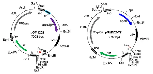
Fig. 5 Bifunctional Streptomyces vectors. Plasmids are based on the minimal replicon of pSG5.