Life Cell Imaging
Due to the triumphal course of autofluorescent proteins such as the green fluorescent protein (GFP), distinct compartments of living plant cells can be fluorescently labelled individually.
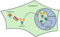
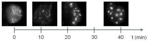
Live Cell Imaging. Left: schematic light dependent activation & transport of phytochrome A (phyA) into nucleus, forming nuclear bodies (NB). Right: Nuclei of transfected tobacco leaves show the time dependent morphological change of NBs (phyA labelled with GFP).
As fluorophores can serve as a sensitive sensor of their immediate chemical environment, we use fluorescence lifetime imaging microscopy (FLIM) techniques to monitor changes in the fluorescence lifetime of these label dyes or variations of the spectral shape. This way, we are able to investigate cellular processes on a molecular level. For example, changes in the fluorescence lifetime point towards a change in the refractive index of the surrounding medium, e.g. the cytoplasm, or to mechanical stress due to conformational changes. Diversities of the spectral shape of a fluorescence marker on the other hand indicate e.g. a change in the local pH. These methods are also incorporated in FLIM-FRET (Förster resonance energy transfer) studies, which we use to elucidate protein-protein interactions in living cells and thereby their function in complex signaling networks.