New microscopic methods
Though we mostly examine biological samples, we benefit from our background in physical chemistry to develop novel microscopic methods, improve current techniques or update our custom-made microscope setups with current state-of-the-art extensions. By this, we can provide highly versatile analytical methods to approach a large scope of research questions - e.g. in vivo measurements of redox potentials in plant cells, distribution of chemical stent coatings into arterial tissue, excitation spectroscopy of single emitters, and in-depth single molecule analyses of fluorescent proteins.
One example for a newly developed method in our lab is fluorescence intensity decay shape analysis microscopy (FIDSAM) which exploits shape differences of fluorescence lifetime decays collected in FLIM experiments to suppress undesired contributions to fluorescence intensities like autofluorescence in plant cells.
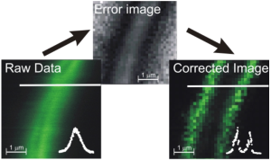

FIDSAM. Left: Enhanced image contrast of Arabidopsis cells GFP-labeled at the BRI-1 membrane protein. After contrast enhancement correction by FIDSAM, the plasma membranes can be optically resolved. Right: both uncoated and coated (reporter gene + GFP) vessel stents show similar fluorescence intensity distributions in raw data images. FIDSAM showed that the stent coating is actually diffusing into the tissue, otherwise concealed by the strong autofluorescent background.