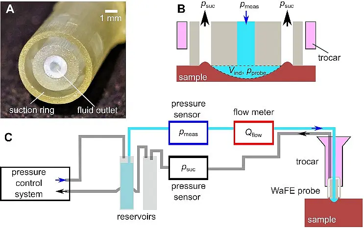
We address questions of biological and medical relevance using physical methods. Our main tools are various species of scanning probe microscopes. Seeking to descend deeper and deeper into the nanoworld, we place the development of new instruments and methods with high-speed and low-noise characteristics at the foundation of our research. Our applications include visualizing the dynamic interactions of single biomolecules, revealing morphological and mechanical properties of living cells and tissues, exposing biomaterials on the molecular scale, and illuminating transport processes in artificial and natural membranes. We also develop elastography methods for tissue differentiation in minimally invasive surgery.
Contact
Prof. Dr. Tilman Schäffer
Institute of Applied Physics
University of Tübingen
https://www.bioforce.uni-tuebingen.de/en
Topics
NanoAnalytics, nanomedicine, mechanics of cells and tissues, elastography, sensor technology, surfaces, scanning probe microscopy, single molecule force spectroscopy.
Atomic Force Microscopy (AFM)
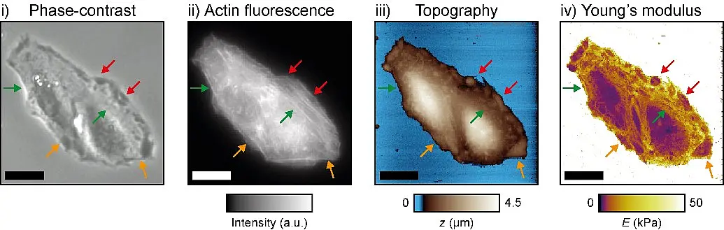
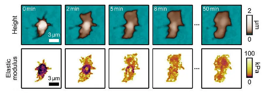
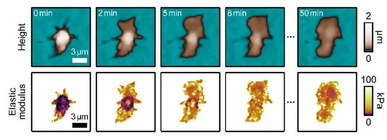
One of our goals is to map dynamical and mechanical properties of structures at the molecular, cellular, and tissue level. For this purpose, we are continuously improving scanning probe microscopy instrumentation and methods. For example, we are improving resolution and measurement speed by designing and building atomic force microscopes for small cantilevers. These small cantilevers have mechanical resonance frequencies in the megahertz range and can be used to image dynamic interactions between single biomolecules in real-time (e.g., protein-DNA interactions), or the complex mechanics of living cells.