Excellence Strategy

Radiooncoclogy

Radiotherapy is a cornerstone of cancer treatment; around every second tumor patient receives radiotherapy during the course of the disease. The research focus of the Department of Radiation Oncology is on personalized, high-precision radiation therapy of tumor diseases. Through translational research in the interdisciplinary team of radiation oncologists, physicists and biologists, the aim is to further personalize the application of radiotherapy using the latest medical technology innovations and developments as well as through the identification and establishment of biomarkers and functional imaging.

MRI-adaptive radiotherapy (MRI-linac)

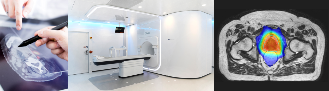
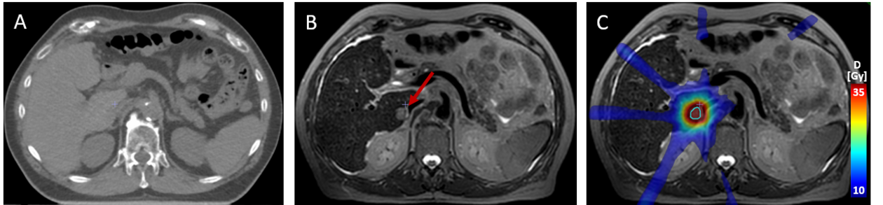
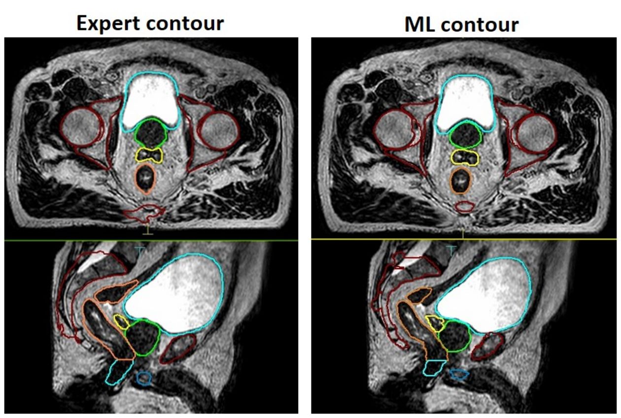
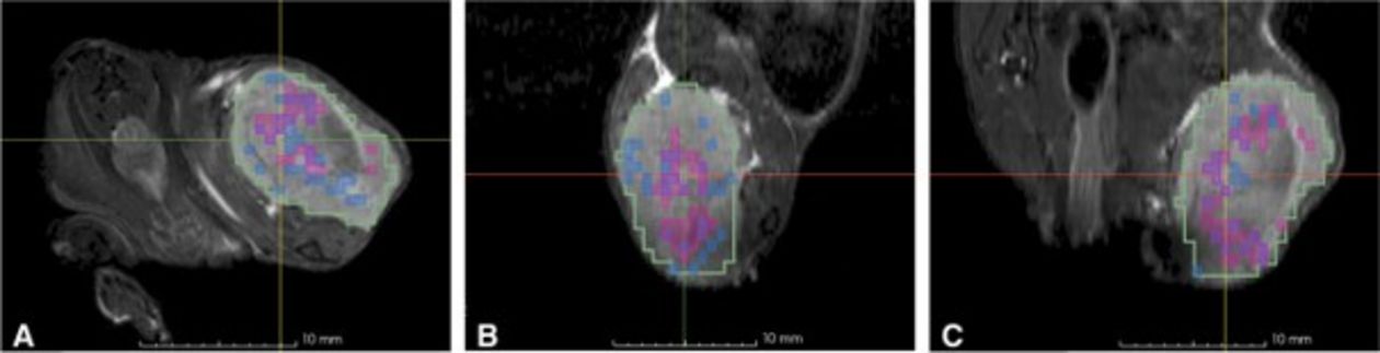
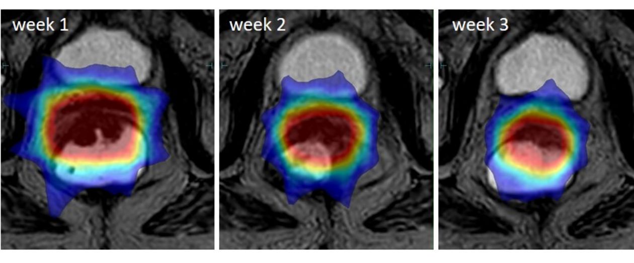
The success of radiotherapy depends on precision. The integration of magnetic resonance imaging (MRI) with a linear accelerator (linac) in a single hybrid device (MRI-linac) for MRI-guided adaptive online radiotherapy is one of the most important technological advances in radiotherapy. During each treatment session, an MRI is performed, the contours of the tumor and the organs at risk are adjusted and the radiation plans are re-optimized. Anatomical changes (tumor growth or shrinkage, edema of healthy tissue) can be detected during radiotherapy, allowing an accurate assessment of treatment-related side effects. Adaptive online MRI not only improves geometric high-precision treatment, but also captures functional and quantitative MRI data, enabling sequential monitoring of quantitative imaging biomarkers (QIB) during radiotherapy. This may enable early response prognosis and individualized dose prescription based on treatment response and biological prognostic factors. The aim of this research focus is to analyze the online integration of anatomical and functional imaging in adaptive radiotherapy.


MR-guided high precision radiotherapy of a liver metastasis. (A) Native planning CT; target delineation is not possible without implanted markers or contrast agent injection. (B) Excellent visualization of the target region using navigated T2-weighted MRI (red arrow). (C) Markerless MR-guided stereotactic body radiotherapy of a liver metastasis using the 1.5 T MR-Linac (Unity, Elekta AB, Sweden) 09/2020 at the University Hospital for Radiation Oncology Tübingen.

Methods of machine learning and artificial intelligence in medical physics and radiation oncology

As part of innovative research projects, the Section for Biomedical Physics is carrying out various aspects of the use and further development of machine learning (ML) and artificial intelligence (AI) in the field of medical physics and radiation oncology. In cooperation with the Cluster of Excellence Machine Learning and the MiDAs Lab of the Department of Radiology, for example, ML methods for the automatic annotation of multi-modal image data are developed and validated for subsequent use in radiation therapy planning. Other research projects deal with the rapid, AI-based calculation of radiation dose in tissue or with the automation of radiation planning by using deep neural networks.

Publications


Exemplary MR-image of a prostate cancer patient with an expert annotation of organs (left) and an automatic AI based annotation (right). The different colors depict specific organs. Nachbar M et al., Z Med Phys 34 (2024) 197–207.

Quantitative MR imaging for radiotherapy

Modern imaging techniques such as positron emission tomography (PET) or magnetic resonance imaging (MRI) not only allow the body's anatomy to be displayed, but also enable the recording and quantification of physiological processes in the human body using functional imaging. The information obtained from this is used in the Section for Biomedical Physics to individualize radiotherapy for patients. PET and MRI imaging are used to better estimate tumor size and to predict patient response to therapy. Another goal is to use functional imaging techniques to identify patient-specific, radioresistant tumor areas that are treated with an increased radiation dose in clinical studies.

Determination of potentially radioresistant tumor areas by calculating 1D (blue) or 3D (purple) clusters from diffusion-weighted MRI and FMISO-PET in preclinical head and neck tumor mouse models. Boeke S et al., Eur J Nucl Med Mol Imaging, 2023 Aug;50(10):3084-3096.

Translational clinical research

The Department of Radiation Oncology conducts studies that combine maximum geometric precision through technological advancements and biological precision. Rectal cancer is one example of this. As part of DFG-funded projects, a new treatment plan for dose-escalated radiotherapy of rectal cancer was established, which leads to complete tumor regression in many patients with early tumor stages. Functional imaging also plays a central role here, acquiring longitudinal data during treatment and evaluating them quantitatively. In the previous CAO/ARO/AIO-16 study on organ preservation in rectal cancer, RNA sequencing was used to identify potential target genes for radiosensitization, which are currently being investigated in more detail as part of research projects.

Example of online-adaptive dose-escalated irradiation of a rectal carcinoma.

Molecular radiation oncology

The currently achievable effectiveness of radiotherapeutic intervention is limited e.g. by the degree of intrinsic resistance of tumors to the effects of radiation (radioresistance). Dr. M. Orth's research group is working on the identification and characterization of biomarkers for such resistance (for example in rectal cancer) and on the preclinical investigation of novel agents directed against identified biomarkers (so-called "targeted agents/drugs") with regard to their potential to enhance the effectiveness of radiotherapy (currently for example in glioblastoma).