Werner Reichardt Centrum für Integrative Neurowissenschaften (CIN)

Networks

Neuronal Networks and Behaviour

This level of observation is concerned with the question of how interconnected populations of neurons interact to generate function. A significant part of this endeavour is closely connected with the question of how neuronal networks generate behaviour, or in case of disease, what aspect of neuronal population activity generates maladapted behaviour. Experiments to solve these questions are conducted mainly in trained waking animals or in humans. Other questions concern the connectivity and specific aspects of information transfer between neurons or groups of neurons. These latter questions typically lead to experiments using histological methods, or to the functional assessment of neuronal activity in either anaesthetized animals, or in vitro preparations (see: Cells) such as brain slices or cell cultures. Theoretical neurobiology (see: Computational Neuroscience) at the level of networks provides the mathematical concepts with which information about the environment or the subject’s behaviour that is carried by population activity, can be formally described and analysed.

Methods

The methods of analysing neuronal population activity can be divided into approaches that are not able to provide single neuron resolution, and others that do. The first class comprises rather old, but still highly useful methods, such as the electroencephalogram (EEG), and its modern complement, the magnetoencephalogram (MEG), both non-invasive approaches amenable to studies of the human brain. The analysis of invasively recorded electrocorticograms and local field potentials (LFP) is akin to traditional EEG, however, in offering a substantially higher temporal and spatial resolution. Some of the more recently developed imaging methods, e.g. the use of visualization of electrical potentials with voltage sensitive dyes, do not yield single neuron resolution (at least where in vivo approaches are concerned), but they do provide a more detailed picture of the spatial distribution of neuronal activity than traditional electrophysiological methods. The last two decades have seen tremendous progress in our capability to assess neuronal population activity with single cell resolution. The first advance was the advent of multielectrode extracellular recordings that allowed the simultaneous recording of action potentials from a large number of neurons. A more recent development is the advent of high resolution 2 photon imaging. In combination with calcium sensitive dyes, 2 photon imaging is able to yield functional signals from multiple single cells and the neuropil (including axons and dendrites) across an entire population of neurons.

Different types of Networks

Investigations in Humans

Neurons interact with each other over a time range of milliseconds. Detailed study of network activity is only possible through measurement at the adequate temporal resolution. Therefore, the study of neuronal networks in humans is still the realm of electrophysiology, which reaches sub-millisecond precision. For EEG recordings subjects are equipped with a set of electrodes (figure, right) that touch the scalp. These electrodes probe an electromagnetic field that is generated by the time-variant activity of thousands of neurons. The more the polarity of a neuron’s electrical field is oriented toward the outside of the scalp, the more it will contribute to the EEG signal. Therefore, EEG recordings are dominated by the activity of large pyramidal cells in neocortical gyri whose orientation is perpendicular to the surface, thereby allowing them to contribute electrical dipoles that can be picked up by scalp electrodes. In contrast, pyramidal cells in the walls of neocortical sulci will not contribute greatly to the EEG signal because their electrical dipole is oriented parallel to the scalp. Neurons that reside below the neocortex contribute relatively little to the EEG signal because their dipoles show little net orientation and, moreover, they are far away from the electrodes. MEG (figure, left) records the same electromagnetic signal but taps the magnetic component rather than the electrical one. As the two are orthogonal, the MEG signal mainly reflects neuronal activity in the wall of the neocortical sulci. Thus, EEG and MEG signals originate from complementary sets of cortical neurons.

Investigations in Animals

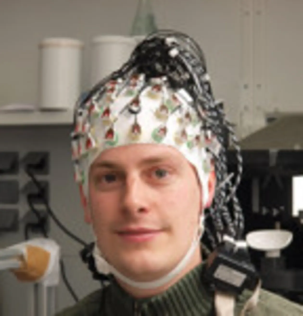
In order to understand neuronal population activity and to understand how it generates behaviour, the activity of each of the contributing neurons and their interaction must be known. Methods such as EEG, MEG, and fMRI that are regularly used in the cognitive neurosciences do not provide the necessary resolution. The fMRI signal is rather indirectly coupled with the activity of rather large groups of neurons, as it essentially measures signals based on changes in brain blood perfusion that are correlated with neuronal activity, but have completely different spatial and temporal properties. Others, such as EEG and MEG, lack the necessary spatial resolution, as they report the sum of the activity of large populations of neurons. The invasive measurement of signals from single nerve cells in humans is limited to rare occasions in which a well-defined clinical problem, such as the search for the source of a seizure, may require invasive scrutiny of the brain.

Although these spin-offs of therapy-oriented invasive approaches have undoubtedly contributed to our understanding of the human brain, their general applicability is hampered by the indispensable priority of the medical goal, which constrains the time available to do the research. Obviously, the scientist involved is not free to select a target structure of choice, but is restricted to the site to be explored for medical reasons. A final qualification pertains to the fact that the brains studied are those suffering from a disabling disease, processing information in an aberrant manner. Studying single and multineuronal signals in healthy animals is devoid of all these limitations and is the only way to acquire information about information processing by neurons in parts of the brain that usually are not involved in clinical procedures.

For more information about the CIN's use of animals in research, and about the legal, ethical, and scientific ramifications please take a look at the animal research section.

Scientific investigation always works with models that are simple and can be easily understood, or are in other ways advantageous - for instance because they are easily amenable to experimental control and manipulation. Using these models, basic insights can be gained which then lead to a more complete understanding of complex models. This is exactly the role of animal models in life sciences. If a suitable model of a human brain system is chosen, the conclusions suggested by study of the model are in most cases valid for the human system at stake.

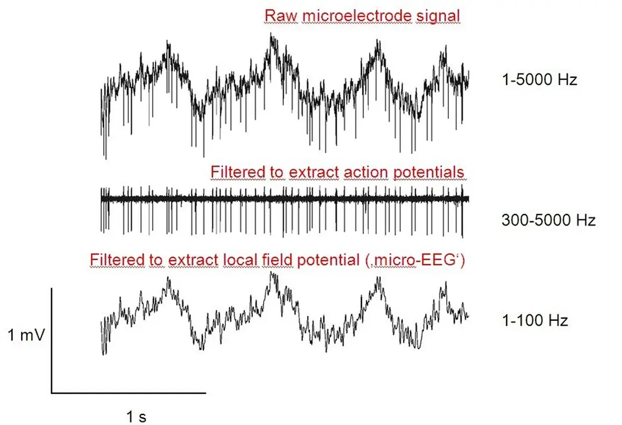
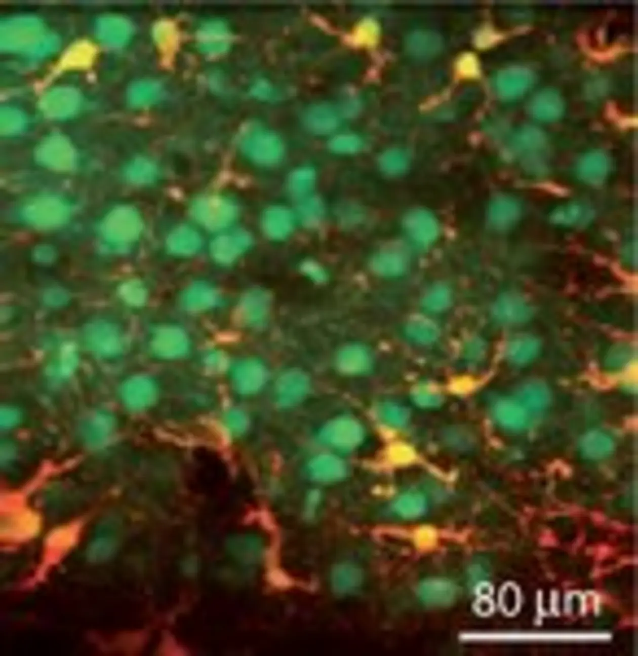
Nevertheless, careful consideration is always warranted and investigators will try to test critically the applicability of a concept by formulating through non-invasive experiments that address the human system, designed on the basis of insights derived via animal experimentation. Animal experiments allow the recording of neuronal signals with unrivalled precision. Small arrays of microelectrodes can be introduced to record from several single nerve cells in parallel without destroying tissue. Furthermore, 2 photon imaging is currently developed to measure complex intracellular chemical signals from multiple single nerve cells at the same time.

Surgery and Behavioural Training

In an aseptic surgery under general anaesthesia, arrays of microelectrodes are introduced to specific sites in the brain of experimental animals such as mice, rats or macaque monkeys. The location of the electrodes is determined by stereotactic coordinates and/or by functional assessment of the response properties of the neurons found. The electrodes are connected to wires and microplugs that are embedded in the head implant. In addition, a head post is implanted into the acrylic. Post-operative care provides analgesics, warmth and the administration of liquid if necessary. Surgery is followed by a recovery period of several weeks in which the wound heals completely. In monkeys, the electrode position within the brain is routinely checked using standard MRI images.

In many neurobiological experiments, the animals are head-fixed and are required to solve quite difficult tasks. For instance, they are asked to discriminate sensory stimuli at their perceptional threshold. To accomplish difficult cognitive tasks, great care has to be taken that the animals are healthy, feel well and experience no stress. Head-fixing has to be introduced in small steps in order to accustom the animal to the experimental procedures. It is absolutely possible to accustom animals to head-fixing without having them struggle or show any aversive reaction. The trick is the same as that used by all animal trainers, whether in science or in a circus: be very patient, introduce potentially stress-evoking situations in very small steps, repeat them often, and reward the animal for successful behaviour. In order to adapt a rat to head-fixing, some four weeks are needed; monkeys take about the same time. The videos provided by our partner institution, the Max Planck Institute for Biological Cybernetics, give excellent insight into animal training for experimentation (see: here).

Once the animals are fully accustomed to the procedure of head fixing, they are trained for the actual task. Again, a complex procedure is learned step by step and each successfully accomplished step is rewarded. Food and drink are the most common rewards in animal training. To use them as rewards, one has to limit the animals’ free access to foodstuff. This does not mean that animals must suffer thirst or hunger. Rather, the foodstuff becomes available only after successful accomplishment of a task. The trainer can adjust the availability of the reward by designing the difficulty of the task or by varying the amount of reward given each time a successful action is performed.

It is a rule of animal training to balance difficulty of the task and volume of single rewards such that the animals acquire enough quantities of foodstuff to reach satiety. Whenever a task is performed well by the animal, a small new step is introduced that makes it a little more difficult to assess the reward and motivates the animal to learn this next step.

Recording Action Potentials (Spikes) and Local Field Potential

Electrical signals from single neurons are typically picked up by microelectrodes that are introduced into the brain. The standard format is a sharpened micro-wire of diameter of less than 100 µm at the shank and less than a 1um at the tip, made from platinum, tungsten, steel, iridium or alloys. The shaft of these wires is insulated electrically and exposes only a very small area of metal at the tip of the wire to the tissue. These active zones are typically smaller than the cell bodies of neurons and thus can be brought close enough to the neurons to pick up signals from just one neuron.

To record many neurons simultaneously many of these electrodes can be assembled into arrays. A more recent variant of multiple micro-electrodes is based on silicon wavers. These hold conducting paths that end in exposed disks, the active zones, which can be machined as small as 10-20 µm in diameter and may have distances as small as 20 µm between each other. Electrical signals recorded against a reference electrode are adequately filtered to isolate the action potentials of neurons that have their main power in the frequency range above 300 Hz. The shape of the extracellularly recorded action potential is determined by the shape of the neuron and the distance and relative location of the electrode. Thus each neuron surrounding the electrode contributes its own characteristic action potential wave-form. This is why these action potentials can be sorted and assigned to single neurons.

Local field potentials are recorded with the same electrodes but considering only low frequency signals between 1-100 Hz. In these frequency ranges, slower membrane potentials originating from synapses or intrinsic voltage gated membrane properties are picked up. In this mode a sum potential of the entire neuropil – a net composed of all the fibres and appendages of neurons - surrounding the electrode is recorded. No single cell resolution is possible in this frequency range.

Recording Calcium Signals Using 2-Photon Microscopy

When neurons become electrically active, the influx of calcium ions is induced through their membranes. This fact is used by calcium dyes to signal neuronal activity. Calcium dyes are fluorescent chemical compounds that respond to excitation with light by emitting light themselves. The colour of the emitted light will vary depending on the concentration of calcium ions in the vicinity of the dye. These dyes are injected into neuronal tissue using a micropipette. Once inside the tissue they pass the cell membrane of neurons and end up inside the neuron, which is where calcium concentration changes constantly with the neuron’s electrical activity. The dye is then excited with the so called two-photon technique.

In a nutshell, two-photon excitation uses two light particles (called photons) of low energy rather than one photon of high energy to excite the calcium dye. The advantage of light at low energy is that it is less scattered and absorbed by biological tissue, and thus it can penetrate deeper into the brain and is less phototoxic than light of higher energy. Another advantage is that two-photon excitation only happens if sufficient numbers of photons are present at the same spot. Thus, while single high energy photons can excite the dye at any (unspecified) point of their trajectories through the tissue, the low energy photons can excite the dye exclusively at the place where there are other photons in sufficient numbers to combine their energies to do the job. By using pulsed laser light and focusing it with a lens, one can thus determine very precisely at which spot inside the tissue excitation will happen and thus from which point a measure of neuronal activation is taken.

Two photon calcium imaging has been developed in anaesthetized animals in vivo but it is on the verge of being applied successfully in waking animals that have been trained. Important precondition for imaging in trained animals is the development of calcium dyes that are non-toxic or and/or are genetically programmed and transfected by a virus, and thus allow repetitive imaging.