Werner Reichardt Centrum für Integrative Neurowissenschaften (CIN)

Methods

Methods in Integrative Neuroscience

Neuronal function underlying behaviour and cognition is organized at many different levels: from genes and molecules to cells, to neuronal networks and large scale neuronal systems involving distinct brain nuclei and regions.

These levels of organizations interact to generate emergent properties on the next higher level. Only detailed knowledge about this interaction lets us fully appreciate the brain's functionality, and allows us to use this understanding for applications in medicine and technology.


The above schema is a visualization of the levels of organization studied by the community of researchers within the CIN. The final goal is to use this knowledge for human applications, typically in medicine as well as for the advancement of modern technologies such as information technology, robotics and artificial intelligence. The graphic also highlights the important role of theoretical neurobiology in guiding experimental work at all levels of organization. Finally, the endeavour as a whole must be accompanied by a philosophical analysis of concepts and paradigms, as well as by ethical considerations.


Overview of the methods

Levels of Organisation

All of biology starts at the level of genetics. In the past there have been high hopes that seemingly complex phenomenons might turn out simpler than expected if they could be traced back to individual genes. However, it turned out that the idea that all complex brain function or neurological disease can be directly and causally related to individual genes is not realistic. The reason is that the diseases where it is theoretically possible to ‘repair’ a single gene and thereby heal the patient are a tiny minority among all known brain diseases. No complex brain function has been found that is caused by one single gene. The reason is that most neuronal functions and dysfunctions are not ‘caused’ by genes in the strict sense. Rather, complex networks of genes ‘predispose’ the brain to function or dysfunction in certain ways. Theoretically, predicting exactly the expression of neuronal (dys-)function requires the knowledge of the exact composition of a vast pool of genes as well as the contributions of environmental factors.

Sets of genes affect whole networks of molecules and cells on the higher levels of organisation in ways as yet unknown, their impacts moving all the way up to cognition and behaviour. Therefore, if neuroscientists wish to understand what is going on when complex brain function is generated, they must undertake the arduous task of finding out how function is generated on each level of organisation and how it is transferred to the next higher one. This understanding rests on the insight originally expressed by David Marr and Werner Reichardt. It is now the mission of the CIN.

Level-Specific Methods

At each level of organisation, CIN investigators apply a variety of tools that allow them to dissect specific aspects of function.

They make use of methods that are focused on the typical scale of each level of organisation and its particular requirements. The first and most important methodical consideration is resolution: whether one employs this or that tool depends heavily whether one wishes to investigate phenomena in the micrometer, millimeter, or centimeter range, as well as the millisecond, second, or minute range.

The following three graphics show the spatial (ordinate) and temporal (abscissa) resolution of the neurobiological tools that are most commonly used. The degree of resolution that is roughly feasible with each of the tools is demarcated by the coloured boxes. Note the logarithmic scale.

Electrophysiological Methods

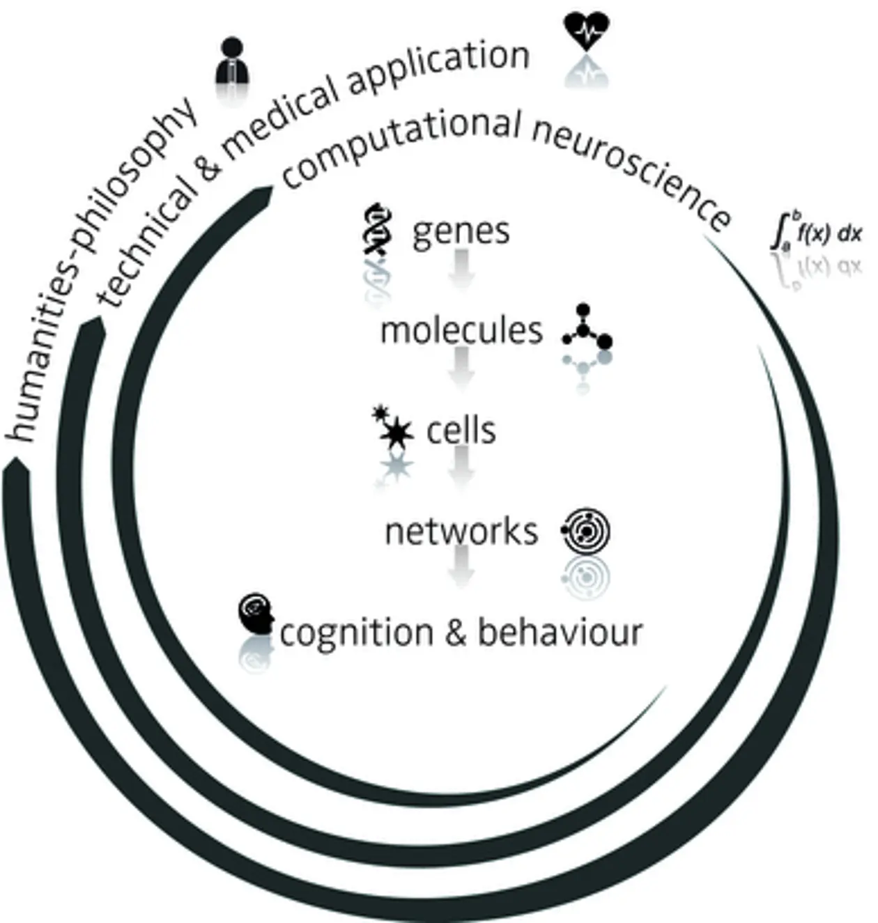
Among the methods that are applicable in studies of the human brain, the electrophysiological methods EEG and MEG have excellent temporal but rather poor spatial resolution. Electrical activity up to 100 Hz can be resolved (millisecond resolution) but the spatial specificity is at the level of the whole brain or sometimes of the cortical lobe.

Modern imaging techniques are better at locating brain activity, but suffer severe drawbacks in terms of temporal resolution. Neither functional Magnetic Resonance Imaging (fMRI) nor Positron Emission Tomography (PET) report neuronal activity on a scale better than a few seconds (see: Cognition & Behaviour). Similar constraints are also valid for employing lesions, which are the most traditional means of studying human brain function. Lesions due to disease (strokes or tumours) usually affect large brain areas and are only rarely small and circumscribed enough to allow conclusions to be drawn about the function of a single defined brain structure or neuronal pathway.

Time-honored research

Electrophysiology using microelectrodes is in many ways still the gold standard of modern neurophysiological research. It has excellent spatial and temporal resolution (in the millisecond and micrometer range). Intracellular techniques, such as patch clamp recordings, can yield data from subcellular compartments such as dendrites and axons (see: Cells). Extracellular recording yields information about single action potentials from single neurons (single unit). Field potentials sample from small networks confined to the sub-millimeter range. Around the same spatial range of neuronal tissue is activated by electrical micro-stimulation (see: Networks).

Optical Methods

New optophysiological methods use light to probe neuronal activity with high temporal and spatial precision. Intrinsic optical imaging and voltage-sensitive dyes report neuronal activity on the scale of milliseconds. However, they are as of yet not able to resolve single neurons.

Calcium imaging uses indicator dyes that are introduced into single cells or cell populations by different means (e.g. incubation or via microlelectrodes). Thus cellular and sub-cellular resolution is reached with this method (see: Networks).

Optogenetic stimulation uses genetic modification of specified neurons. Here, light-sensitive ion channels or pumps are inserted into the neuron’s membrane. Complex genetic constructs using genes that are transcribed only in specific tissues can be used to specifically label sub-populations of neurons. Stimulation of neuronal subclasses or single neuron stimulation may eventually become feasible with this method.

Other Methods

Last but not least, the methodological toolbox of molecular biology and genetics for assessing the function of molecules is vast (see: Genes and Molecules). These methods permit the measurement or manipulation of the function of single molecules and molecular signalling cascades. These methods can be targeted at single cells as well as at whole organisms. They thus cover measurements over a large range of temporal and spatial scales.