Werner Reichardt Centrum für Integrative Neurowissenschaften (CIN)

Ophthalmic Research

Signal Processing in Retina

Visual information processing begins in the retina, a thin neuronal tissue lining the back of the eyeball. As a part of the brain, the retina does not only convert the incoming stream of photons into electrical signals, it also performs a detailed and highly specific analysis of the observed scene. Therefore, the retina can be considered a highly specialized and sophisticated image processor.

All visual information sent from the retina to the brain travels along the optic nerve, a major bottleneck of the visual system. Therefore, prior to transmission to the brain, important aspects of the observed scene must be extracted and encoded as spike patterns. These features include simple ones such as contrast, brightness and “colour”, but also more complex ones, such as information about objects moving relative to the background. Thus, the retina sends in parallel many representations of the visual scene to the brain; each of these representations encodes different features and is represented by one of the roughly 40 retinal ganglion cell types whose axon form the optic nerve. The importance of retinal signal processing is highlighted by the fact that important decisions – what visual information is relevant, and what can be safely discarded – is made already in the retina.

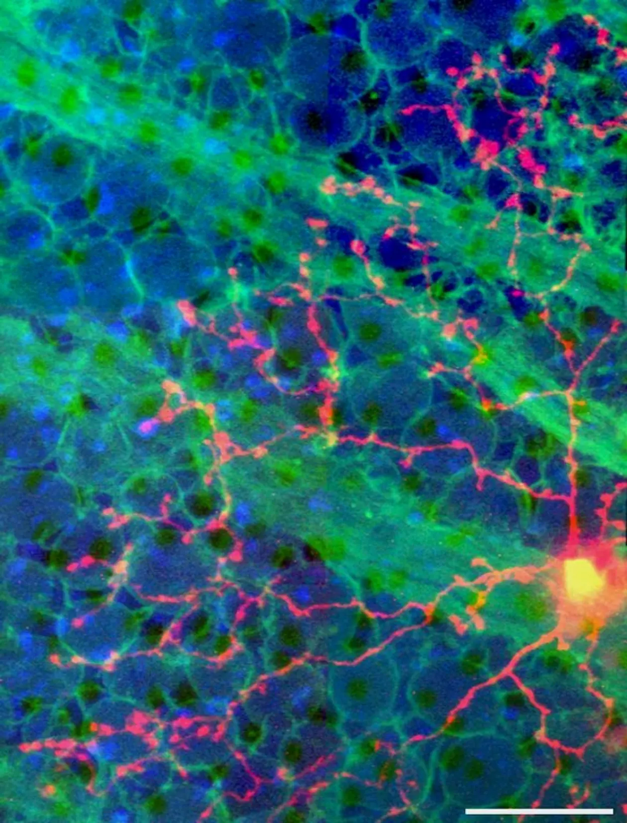
The computational capabilities of this intricate neuronal network rely on nearly 100 types of retinal neurons organized in complex microcircuits. Our work aims at unravelling function and organization of retinal microcircuits towards a better understanding of the underlying computational principles. Furthermore, we are interested in how these circuits are altered during degeneration.

Methodology

We established a comprehensive method catalogue for optical measurements of light-driven population activity along the retina’s entire vertical pathway based on synthetic and genetically encoded fluorescent activity sensors.

Our key technique is two-photon microscopy, which enables us to excite fluorescent probes in the intact living retinal tissue using infrared laser light, with minimal effects on the light-sensitive photoreceptor pigment. Therefore, we can simultaneously record activity in neurons at both population and subcellular levels while presenting sophisticated light stimuli. This approach is complemented by single-cell electrophysiology and immunocytochemistry, as well as large-scale data analysis in close collaboration with the groups of Philipp Berens, Matthias Bethge, and Katrin Franke at Tübingen University.

Research questions

  • Local circuits – How do individual neurons at different stages of the retinal network process information in their dendrites and/or axon terminal systems?
     
  • The retinal code – How are the numerous parallel output channels that are present at the level of the ganglion cells set up in the retinal network? What visual features are encoded in these channels?
     
  • Visual ecology – To what extent is the mouse retina adapted to the animal’s natural visual habitat? What functional roles do retinal specializations - such as the opsin expression gradient - fulfill in this context?
     
  • Health and disease – How does the retinal network rewire and alter its function when photoreceptors degenerate?

Collaborations

  • Tom Baden, University of Sussex, Brighton, UK
  • Philipp Berens, CIN / Institute of Ophthalmic Research, University of Tübingen, Germany
  • Matthias Bethge, CIN / Institute for Theoretical Physics, University of Tübingen, Germany
  • Kevin Briggman, Caesar, Bonn, Germany
  • Karin Dedek, Dept. of Neurobiology, University of Oldenburg, Germany
  • Winfried Denk, MPI of Neurobiology, Martinsried, Germany
  • Katrin Franke, BCCN / MPI biol. Cybernetics / Institute of Ophthalmic Research, University of Tübingen, Germany
  • Silke Haverkamp, Caesar, Bonn, Germany
  • Andrew Huberman, Dept. of Neurobiology, Stanford University School of Medicine, CA, USA
  • Markus Meister, Caltech, Pasadena, CA, USA
  • Sebastian Seung, Princeton Neuroscience Institute and Computer Science Dept., Princeton, NJ, USA
  • Robert Smith, Dept. of Neuroscience, University of Pennsylvania, Philadelphia, PA, USA
  • W. Rowland Taylor, University of California Berkeley, CA, USA
  • Rachel O. Wong, Dept. of Biological Structure, University of Washington, WA, USA