Uni-Tübingen

Research Stay Boston, USA 2023

Doctoral Fellow: Tamara Hussein (2nd Cohort)

Tamara Hussein (P6-neuroprotection) joined the lab of Professor Michele Jacobs' at Tufts University Graduate School of Biomedical Sciences.

The research group focusses on exploring human gene mutations associated with intellectual disability and autism, with a specific emphasis on elucidating the pathophysiological alterations across behavioural, molecular, synaptic, and circuit levels. These mutations are anticipated to interfere with the adenomatous polyposis coli protein, β-catenin, and the canonical Wnt- signalling pathway.

During her research stay she was trained to implement various biochemical methods but primary focus was directed on performing electrophysiological measurements on ß-Catenin deficient mice to elucidate synaptic functionality. Utilizing extracellular recordings, she investigated the excitatory postsynaptic potential (EPSP), reflecting the postsynaptic response of downstream CA1 pyramidal cells. This approach facilitated the exploration of neurobiological mechanisms at the synaptic interface. Systematic manipulation of the frequency and intensity of presynaptic stimuli enabled the analysis of diverse physiological parameters of synaptic transmission. Of particular interest was the investigation of long-term potentiation (LTP), a form of synaptic plasticity recognized as a neurobiological correlate of spatial learning. Investigations into LTP and other synaptic processes significantly enhances the comprehension of the neuronal mechanisms underlying learning and memory.

The skills, expertise and insights gained during her research visit in Boston will significantly enhance her ongoing research in Tübingen. A special thank you goes to the DFG and the Reinhard Frank Foundation for their financial aid for this research stay.

Research Stay St. Louis, USA 2023

Doctoral Fellow: Tom Schwerd-Kleine (2nd Cohort)

In June 2023, I had the opportunity to join the laboratory of Philip Williams at Washington University in St. Louis Medical School, department of Ophthalmology and Visual Sciences for a three-month research project.

The primary long-term goal of the Williams lab is to develop new strategies to improve patient outcomes following CNS injury and neurodegenerative disease. The lab uses the mouse eye as a model system to study neuronal degeneration and regeneration since it provides many unique advantages including ease of accessibility and well documented neural circuit components. In the Williams Lab, I utilised multiphoton microscopy to study the Nitric Oxide/cGMP signalling pathway in retinal ganglion cells (RGCs) of mice in vivo. With this technique, combined with a virally expressed biosensor for cGMP, I could study potential heterogeneity of Nitric Oxide/cGMP signalling in different RGC types. Additionally, I implemented an open source based visual stimulation system which will allow the lab to study light evoked RGC responses in vivo in the future.

Philip and all my colleagues in the lab were very welcoming, supportive, and helpful in introducing me to their techniques and helping me with any questions. Therefore, during my (much too quickly passing) time in the US, I learnt a lot scientifically and made great new friends at lab socials over fun board games, St. Louis food and delicious homemade ice cream.

Also, I discovered a commemorative plate for the Nobel laureate Robert F. Furchgott who contributed to the discovery of Nitric Oxide as a signalling molecule in mammals.

Finally, I want to greatly thank the GRK2381 for funding me for this research stay and also Philip Williams and all his lab for having me there and making my time in St Louis such an unforgettably great experience.

Research Stay Boston, USA 2022- 2nd cohort

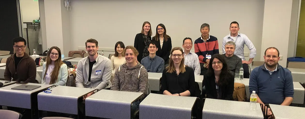
Doctoral fellows: Jennifer Schulz, Aylin Balmes, Timo Kopp, Dila Calis

Research Stay Report

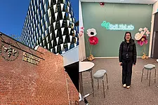
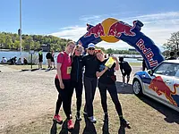
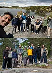
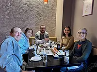
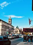
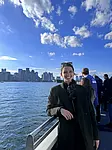
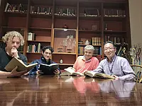
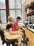
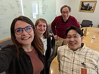
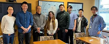
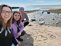
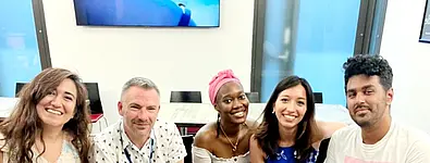
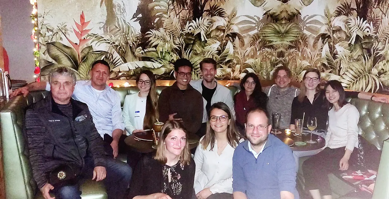
Between April and July 2022 four PhD students from the 2nd cohort of the GRK 2381 in Tübingen had the chance to join different laboratories in Boston for a three-month research visit, just like several students from the 1st cohort did in the months before.

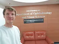
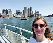
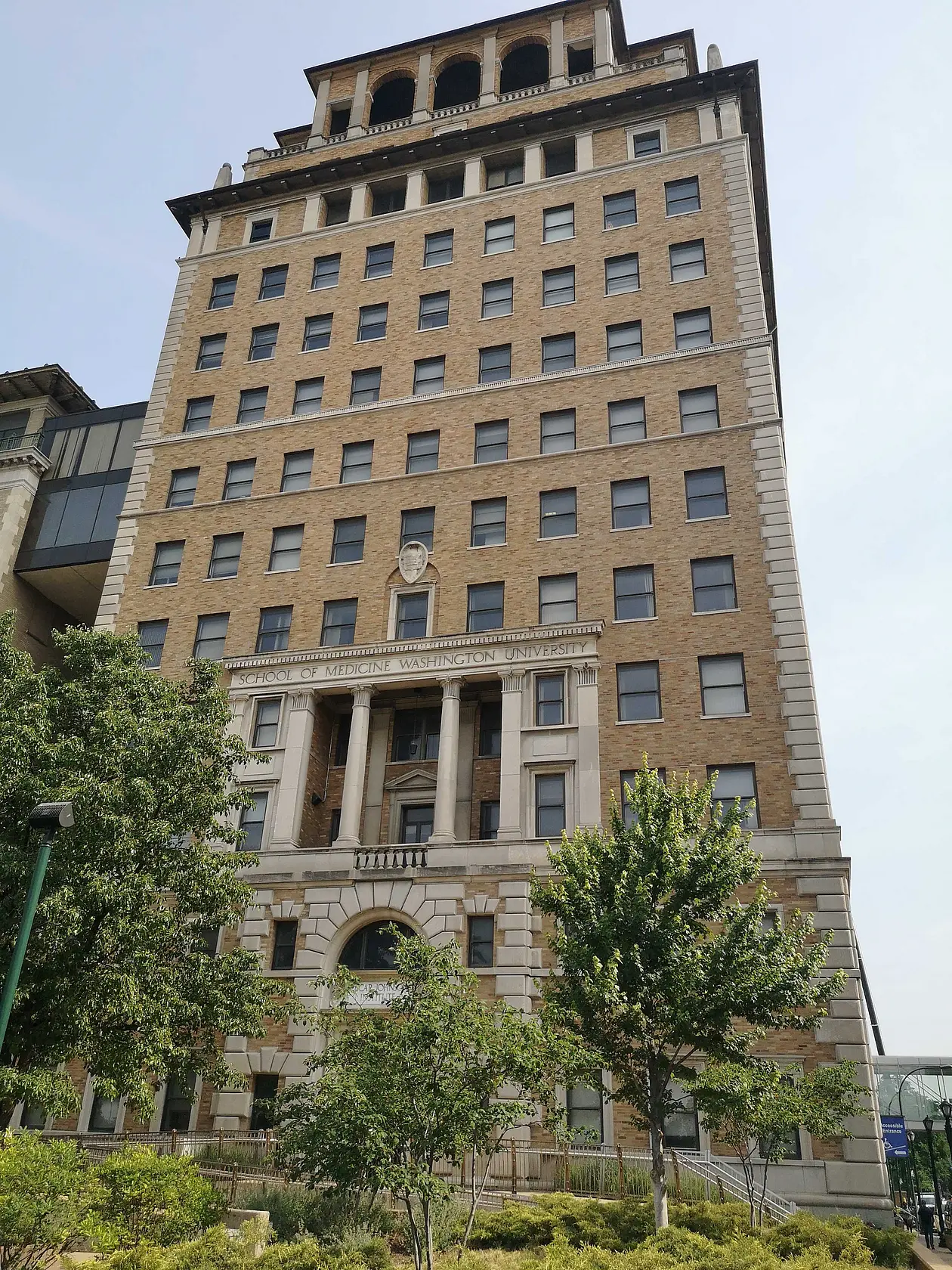
Jennifer Schulz (P1 – cancer) joined the E. L. Steele laboratories for Tumor Biology at the Massachusetts General Hospital and Harvard Medical School. Here, she worked with the group of Associate Professor Dai Fukumura. The focus of the laboratory is to understand vascular, interstitial and cellular barriers to the drug delivery in solid tumors with the aim to discover and test new treatment strategies to overcome the physiological barriers.
During her research stay she investigated, how different drug treatments (in combination with radiation) affect the tumor microenvironment in pancreatic adenocarcinoma (PDAC). She was able to learn and practice various techniques, such as radiation and immune-checkpoint blockade treatment of tumor-bearing mice, which all of them are standard-of-care treatments for patients, offering the potential to translate research results from bench to bedside.
In addition, she expanded her skills in isolating tumor stroma cells through complex flow cytometry panels to reveal how the distribution of certain stroma cells within the tumor microenvironment is affected by various drug combinations.
Furthermore, she was able to improve her knowledge about melanoma mouse models due to the great expertise of the laboratory and the excellent training she received.
Back in Tübingen she will implement the newly acquired techniques in her own PhD project and stay in touch with the great scientific community in Boston.

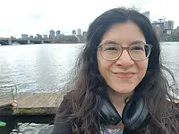
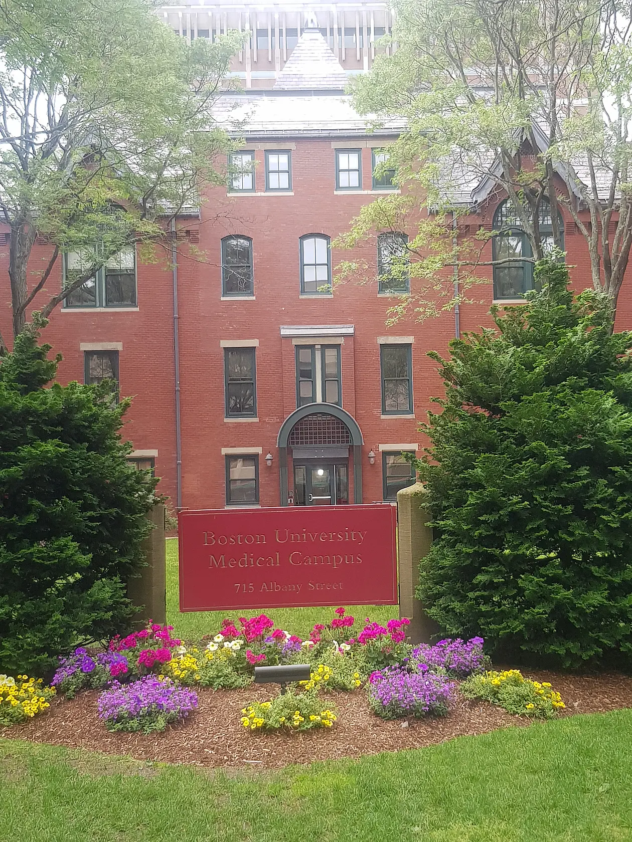
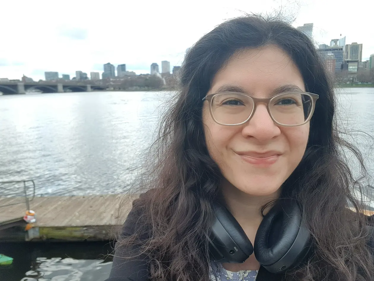
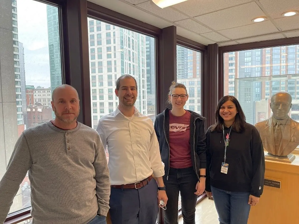
Aylin Balmes (P3 – vascular biomechanics) joined the laboratory of Associate Professor Francesca Seta at the Boston University School of Medicine as a visiting scholar. The main focus of the laboratory is the investigation of basic mechanisms of vascular disease, with a focus on the role of vascular smooth muscle cells (VSMCs) in vascular remodeling.
During her stay she learned how to measure pulse wave velocity (PWV), the gold standard measure of arterial stiffness, in vivo in mice. These experiments are a great complement to her experiments in Tübingen where she studies stiffness on the cellular level using scanning ion conductance microscopy (SICM) and atomic force microscopy (AFM).
During her research visit she investigated if vascular smooth muscle cell stiffness is linked to actin cytoskeleton remodeling. This can be quantified using different techniques. One of those is filamentous (F)-actin immunostaining, which she previously used in Tübingen. Another one is Western Blot quantification of filamentous (F)- and globular (G)-actin, which she learned in Boston. Additionally, she learned a lot about the molecular biology of VSMCs particularly the role of the cyclic guanosine monophosphate (cGMP) pathway.

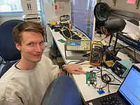
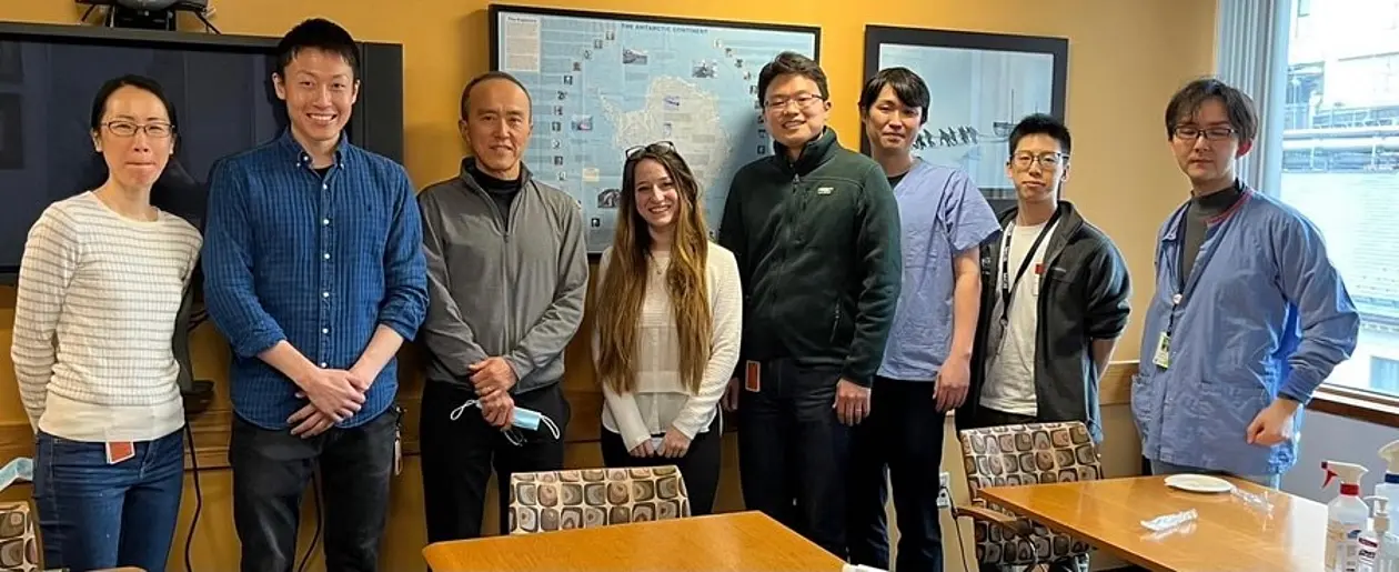
Timo Kopp (P4 – atherosclerosis) joined the laboratory of Associate Professor Dr. Dmitriy Atochin at the Charlestown Navy Yard in Boston to conduct vascular research. The laboratory focuses on ischemic stroke models and possible treatment options for patients experiencing a stroke. Investigating the role of vascular smooth muscle cells (VSMCs) in reperfusion and limiting the damaged area in the brain is of particular interest in this regard.
During his time there, his work focused on assessing the effects that a near-infrared laser has on the vascular reactivity in mice and how that effect might be beneficial for the outcome of stroke experiments. This series of experiments included breeding and genotyping mice of a specific genetic background (wild type, eNOS A-K.I.; eNOS D-K.I. and cGKI K.O. mice), checking for phosphorylation of eNOS and Akt after laser exposure by Western Blotting and most importantly, investigating changes to cerebral blood flow in mice that underwent stroke surgery, after their skull was exposed to the laser.

In addition to this, he was involved in a study about the effects of the same laser setup on a cancer cell line. Observed effects might give an indication that this technique can be used to treat the same cancer in humans as well.
As of August 2022, both projects resulted in co-authorships of two publications in the FASEB journal, underlining the mutual benefits of this research co-operation.

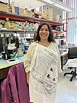
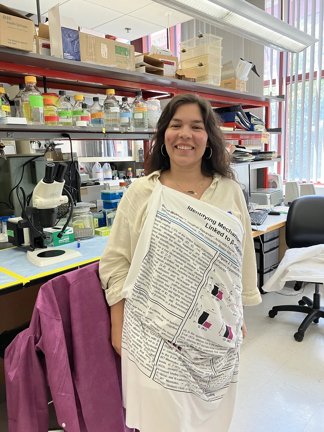
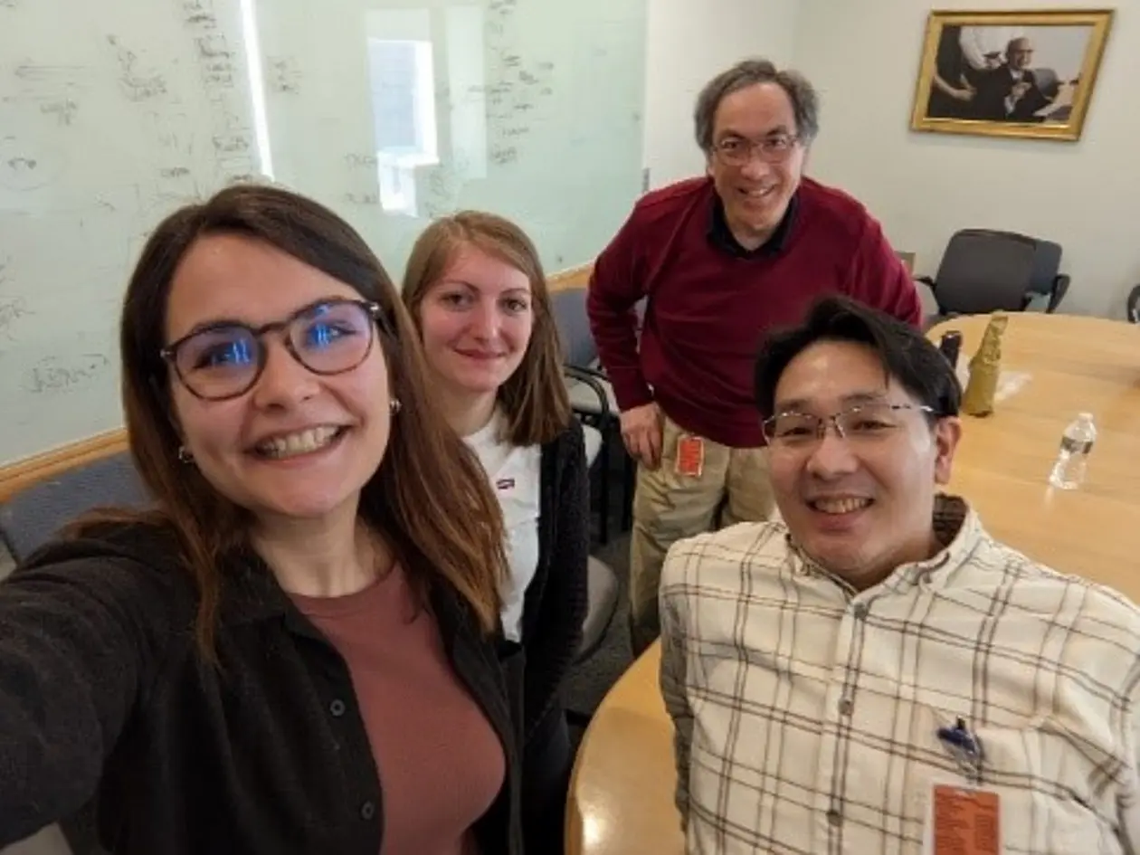
Dila Calis (P9 – brain) joined the laboratory of Professor Michele Jacob at the Tufts University Graduate School of Biomedical Sciences. The research team mainly focus on the Wnt-signaling pathway as an underlying mechanism for intellectual disability and autism spectrum disorder by determining the pathophysiological changes on behavioral, molecular, synaptic, and circuit levels.
During her visit, she was trained on different microscopy methods for imaging pyramidal neurons from hippocampus. For her research question she investigates synaptic spine density and structural morphology changes in hippocampal neuron. Synapses are unit structures that function in rapid information processing. It has been reported that spine density and morphology undergo alteration with hearing loss (either age-dependent or after acoustic trauma).

For this purpose, she used Golgi-Stained mouse brain sections that she prepared from different treatment groups, and acquired high-resolution images. In the next step, she was trained on the image analysis by using RECONSTRUCT software which helps to objectively measure density and classify spines.
Her preliminary findings suggest that, preventing the degradation of the cyclic guanosine monophosphate (cGMP) could be protective against the de-arborization of the neurons in the brain upon hearing loss and could be a potential therapeutic target.
She will establish the protocol that she learnt at Prof. Michele Jacob’s lab in Tübingen where she will use this method in different mouse line (conditional KO) as a part of her PhD thesis.

We are convinced that the skills and knowledge we acquired during our research visit in Boston will be of great value for our continuing research in Tübingen.
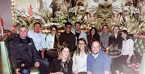
Our gratitude goes to the GRK 2381 and the Reinhard Frank-Stiftung, for providing us with this great opportunity to broaden our scientific horizon and allowing us to connect with scientists from abroad. We would also like to thank Dai Fukumura, Francesca Seta, Dmitriy Atochin and Michele Jacob for allowing us to work at their laboratories and for the great mentorship.

Research Stay Boston, USA 2022- 1st cohort

Doctoral fellows: Marigiovanna Barresi, Alexandra Böttcher, Malte Roeßing, Melanie Cruz Santos, Thomas Pham

Research Stay Report

At the start of 2022, we had the great opportunity to travel to Boston together as a group of five doctoral fellows of the 1st cohort. During this three-month research stay, we could expand our scientific horizons and learn valuable lessons both for our PhD projects and future careers.

Mariagiovanna Barresi (P1 – cancer) joined the E. L. Steele laboratories at the Massachusetts General Hospital and Harvard Medical School. In particular she worked with the team of Associate Professor Dai Fukumura. The expertise of the laboratory in tumor biology allowed her to improve her knowledge in breast cancer mouse models (Jain et al., CSHL Press, 2012). Furthermore, she could expand her experience in cancer research investigating the important role of the tumor microenvironment on drug treatment in pancreatic adenocarcinoma (PDAC).

Alexandra Böttcher (P7 – axon branching) joined Associate Professor Robert Blanton’s team at the Molecular Cariology Research Institute of the Tufts Medical Center. Here, she could improve her experience with biochemical screening methods such as pull-down studies using a cGKI-leucine zipper mutant protein (Calamaras et al., JCI Insight, 2021). Additionally, she introduced an axon labeling technique of Tübingen PI PD Dr. Hannes Schmidt utilizing the lipophilic tracer DiI to study axon branching patterns of sensory neurons in mouse models available in the Blanton lab.

Melanie Cruz Santos (P2 – cardiac injury) joined the laboratory of Professor Fumito Ichinose at the Anesthesia Center for Critical Care Research of Massachusetts General Hospital and Harvard Medical School. During her stay, she successfully expanded the scientific exchange between Tübingen and Boston, as she was trained in a murine cardiac arrest and cardiopulmonary resuscitation model (CA/CPR) (Minamishima et al., Circulation., 2011) and established an acute open-chest in situ model of ischemia and reperfusion injury utilized in her work in Tübingen.

Thomas Pham (P6 – neuroprotection) joined Associate Professor Jesse D. Roberts in his laboratory at the Cardiovascular Research Center at Massachusetts General Hospital and Harvard Medical School. He was trained in wide-field imaging and statistical modeling to localize proteins within sub-cellular compartments, using molecular imaging techniques.

Malte Roeßing (P4 – atherosclerosis) joined the group of Assistant Professor Dmitriy Atochin at the Cardiovascular Research Center at Massachusetts General Hospital and Harvard Medical School. The middle cerebral artery occlusion (MCAO) model (Atochin at al., Neurosci. Lett., 2016) he received training in, can now be established in Tübingen. Malte also worked on multiple other projects, including the study of different therapeutic options available to modulate the NO/cGMP pathway in stroke mouse models.

Next to the amazing scientific experience, we also enjoyed connecting to the international science community in Boston, especially during the Reinhard-Frank-Mini-Symposium. Another cultural highlight for us was cheering for the Celtics and the Bruins on their home turf at the TD Garden Stadium.

We would like to thank all our PIs in Boston and Tübingen for their support in making this research stay a reality for all of us. A special thank you also goes to the DFG and the Reinhard Frank Stiftung for their financial aid

Research Stay Bern, Switzerland 2022

Doctoral Fellow: Lena Birkenfeld (2nd Cohort)

From September to November 2022, I visited and worked in the laboratory of Prof. Dr. Dr. Hans-Uwe Simon at the Institute of Pharmacology at the Inselspital Bern in Switzerland. The research group is working on immunopharmacology and investigates the identification of new drug targets for future therapies in various inflammatory disease models. Further, they are experts in working with immune cells, including neutrophils.

In my project, I am studying the role of neutrophils in the heart after myocardial infarction with a focus on the cGMP pathway in these cells. While I am working with immortalized neutrophils or murine primary cells in Tübingen, my 3 months stay in Bern allowed me to learn the isolation of human blood neutrophils. With this cell type I was able to validate the translational potential of my murine neutrophil data.  In addition, I applied cGMP pathway modulating drugs to investigate their effect on different defense mechanisms of human primary neutrophils using flow cytometry and other techniques as readouts.

Not only in terms of work, but also on a collegial level, it was very nice at the lab in Bern. I quickly found my way into the group and was very warmly welcomed and rapidly integrated. The supervision was also great and I was never left alone with questions or problems.

Besides my stay in the lab, I used my free time to explore the city and get to know the beautiful Swiss landscape and its people.

I would like to thank the GRK 2381 for enabling and financially supporting this research stay abroad. During these 3 months, I was able to generate new and exciting data for my project. Also, I learned and applied many new techniques and methods, of which some were transferred to Tübingen.

Research Stay London, UK 2022

Doctoral Fellow: Johanna Rodriguez (1st Cohort)

In May 2022, I had the opportunity to visit and to work in the lab of Dr. Mike Emerson at the National Heart and Lung Institute at Imperial College London in United Kingdom.  The lab of Dr. Emerson has a strong expertise in Platelet and Cardiovascular Biology. I worked on a project we developed collaboratively to extend my PhD studies to a more clinical setting, including exploration of platelet function and the role of NO-GC/cGMP pathway in people living with HIV. I gained different hand-on experience in platelet and endothelial cell biology including flow cytometry, endothelial-microparticle isolation, light transmission aggregometry, and cell culture. I also had the chance to carry out extensive experiments in the Facility in Light Microscopy at Imperial, where I adapted my microscopy experience to my new set of experiments. I also gained experience with clinical research with patients from Chelsea and Westminster Hospital where I had the chance to learn the importance of patient demographics in clinical research. As a result of my internship at Imperial, we were able to jointly author a paper. The work environment in the lab of Mike Emerson was very friendly and I integrated very well with the team. Our weekly meetings were an informal opportunity to share our research and to hear everyone´s ideas. I always felt very welcomed by the team and the other research groups in NHLI. More than just colleagues, we became very good friends. Besides, the fruitful research stay at Imperial, London is a very vibrant city with many places to visit and activities to go. I would like to thank the GRK 2381 for providing financial support for my research stay in the lovely city of London, where I have the most precious memories that I will always cherish.

Research Stay Paris, France 2022

Doctoral Fellow: Dominic Gonschorek (1st Cohort)

In March 2022, I had the opportunity to visit and work in the lab of Dr. Olivier Marre at the Institute de la Vision, Paris in France for about 3 months. The lab of Dr. Olivier Marre wants to understand how a network of neurons together can code visual information and be the basis of our perception.

In my PhD. project, I want to understand how the neuromodulator nitric oxide (NO) affects the visual signal processing in the inner mouse retina by modulating the second messenger cGMP. Specifically, I research how the output from the retina via the various types of retinal ganglion cells (RGCs), which encode several features from the visual environment, is modulated by cGMP. Therefore, I record the visually-evoked activity in RGCs via two-photon calcium imaging and analyze their activity using machine learning tools.

In the lab of Dr. Olivier Marre, I learned another technique to record neural activity of RGCs, namely multi-electrode recordings using multi-electrode arrays (MEA), which contain up to thousands microelectrode. During my research stay, I performed several experiments using the NO donor DETA/NO and sGC antagonist ODQ.

The MEA has the advantage to directly record spiking activity of RGCs with high temporal resolution as well as to record over a long period of time (since there is no photobleaching caused by the laser), yet, its spatial resolution is fairly low compared to two-photon imaging. Also, identifying specific cell types is more challenging, however, recording activity over a long duration enables to identify RGC receptive fields (RFs), i.e., the sensory space that can elicit neuronal responses when stimulated. These RFs provide important information about RGC spatial-temporal features. Since estimating RFs is difficult with two-photon imaging due to the limited recording time, combining these two datasets, will help us to obtain valuable insights about the temporal-spatial features of the effect on NO-induced cGMP modulation in RGCs.

One highlight of my research stay in Paris was that we recorded from mouse lemur retinae. Close to Paris, there is a mouse lemur colony, which has been able to breed them since the 70s. The special aspect about mouse lemurs is that they are the smallest living primates with a total body size of less than 27cm.

The working environment in the lab of Olivier Marre was very vivid and enabled a productive exchange of knowledge. I received a lot of useful input regarding my project and was supported in any possible way. Additionally, I have been able to further develop and improve my analytical and quantitative skills.

Aside the research in the lab, I could enjoy all facets of Paris by visiting many of its museums such as the L’Orangerie and Grande Galerie de l’Évolution. Also, I could learn and practice a little French.

I would like to thank the GRK 2381 for providing financial support for my research stay in the beautiful city of Paris.

Research Stay Montpellier, France 2021

Doctoral Fellow: Philine Marchetta (1st Cohort)

From September to December 2021, I had the pleasure to work in the laboratory of Prof. Dr. Jean-Luc Puel at the Institute of Neuroscience in Montpellier. Prof. Puel leads the research team "Audition", which among others deals with the logic of sound coding.

One of the mouse models I used during my PhD is the BdnfPax2 KO mouse, which has normal "basic auditory function" but reduced precise temporal auditory resolution that affects cognition and behavior (Eckert, Marchetta et al. 2021, Front. Mol. Neurosc.). My aim was to determine the (dys)function of afferent auditory nerve fibers (ANFs) in these animals, which represent the first postsynaptic contact to sensory hair cells in the cochlea, the inner hair cells. These ANFs can be classified in fibers with a high spontaneous firing rate that have a low detection threshold and others with a low spontaneous firing rate and a high threshold – both of them are important for sound coding of the entire dynamic range. While single ANF recording is very complicated in mice and also too invasive for non-final experiments, the Puel-lab constructed a good alternative. Here the neuronal activity was measured at the round window of the cochlea as a peri-stimulus time response (PSTR) (Huet et al. 2022, J. Neurosci). With the help of Dr. Jérôme Bourien and Dr. Artem Diuba, I improved my round-window-surgery skills in mice, learned how to set the measurement parameters to measure PSTR and did the corresponding data analysis in MatLab.

With the PSTR, I learned a valuable technique, which I am now establishing in our lab in Tübingen. In addition, good scientific contacts were made with Montpellier, and of course I enjoyed the beautiful Camargue and learned a little French on the side.

I would like to thank the GRK 2381 for providing financial support for this research stay.